Back Pain After 50
Age-related spine changes, why some things improve and some worsen with age, and the exercise and lifestyle approach for the over-50 spine.
What Actually Changes After 50
The spine after 50 is not simply a more worn-out version of the younger spine. The changes that occur are specific, predictable, and — in some ways — surprisingly nuanced. Understanding them removes the fatalism that prevents many people over 50 from engaging with rehabilitation.
Disc dehydration and height loss. The lumbar discs progressively lose their water content after the third decade. The nucleus pulposus — the gel-like core that distributes load — becomes less hydrated and less capable of equalising pressure. The disc becomes thinner. At individual spinal levels, this height loss can reach several millimetres by the sixth decade. The immediate consequence is a slight reduction in the height of the intervertebral foramen (the opening through which nerve roots exit the spine), which reduces the available space for the nerve.
Facet joint osteoarthritis. The facet joints — the small paired joints at the back of each vertebral level — develop the same cartilage loss and reactive bony changes that affect weight-bearing joints elsewhere in the body. Facet OA produces morning stiffness, pain with sustained extension, and a characteristic ache at the end of the day after prolonged weight-bearing. It is one of the most common structural findings on lumbar imaging after 50.
Ligamentum flavum thickening. The ligamentum flavum is a thick elastic ligament that lines the back of the spinal canal. With age, it becomes stiffer and thicker — particularly in people who have had chronic low-grade spinal inflammation. This thickening is the posterior component of the stenosis problem.
Bone density reduction. Bone mineral density peaks in the early 30s and declines progressively thereafter. In women, the rate of loss accelerates markedly in the years around menopause. Below a threshold density, vertebral fracture risk increases — even from ordinary daily activities.
The Paradox of Natural Stabilisation
Here is a fact that surprises many people: disc degeneration in the later stages of its natural history actually tends to reduce pain rather than increase it.
The early and middle stages of disc degeneration — the 30s through 50s — are the most painful. During this period the disc is breaking down, inflammatory chemicals are present, and movement at the affected segment is excessive and painful. But as degeneration progresses to its later stages, the disc height loss becomes severe enough that the segment naturally stiffens. Osteophytes (bony spurs) form at the disc margins and act as natural fusion material. The movement at the segment decreases.
This is why many people in their 60s and 70s report that back pain which was severe in their 50s has genuinely improved — not because they did anything specific, but because the natural history of disc degeneration has reached its stabilising phase. This is cold comfort for someone in their early 50s, but it is clinically important: the pain trajectory for disc-based pain is not indefinitely upward.
The natural stabilisation of late-stage disc degeneration is not a reason to avoid rehabilitation — it is a reason to rehabilitate actively during the painful decades to maintain function and muscle mass so that when stabilisation occurs, you have preserved the capacity to live fully. People who wait passively for pain to resolve often emerge from the painful decade with significant deconditioning and muscle loss that limits their function regardless of pain reduction.
The Emergence of Stenosis
Lumbar spinal stenosis — narrowing of the spinal canal — is distinctly rare before 50 and becomes progressively more common after it. The anatomy explains why: stenosis requires the combination of disc height loss, ligamentum flavum thickening, and facet OA to occur simultaneously at the same level. This takes decades to develop.
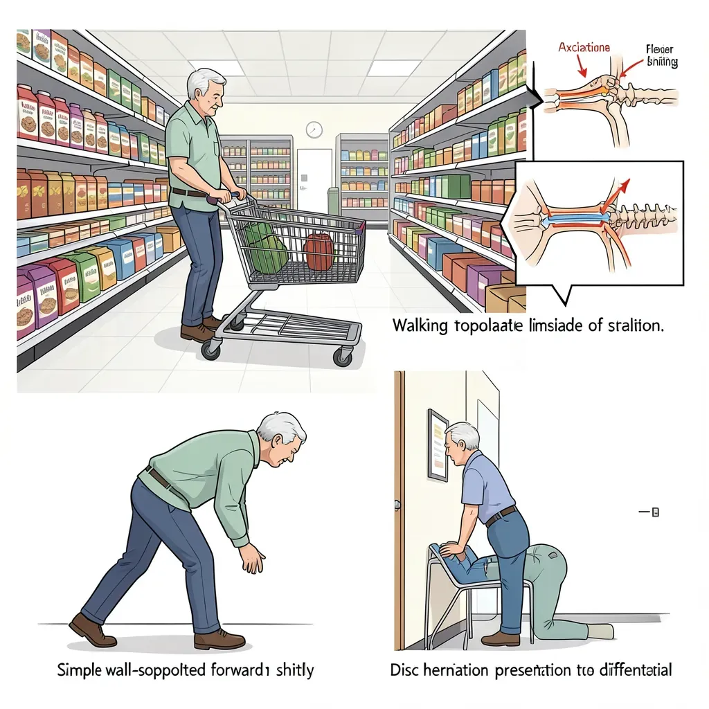
The hallmark symptom is neurogenic claudication: leg pain, heaviness, or weakness brought on by walking and relieved by sitting, bending forward, or leaning on a shopping trolley. The walking tolerance is characteristically limited to a specific distance — 100 metres for some people, 400 for others — before symptoms force a stop. The position of relief is always flexion.
The reason is mechanical: lumbar extension narrows the posterior spinal canal. In a normal spine, this narrowing is trivial. In a stenotic spine, the available canal space has already been reduced by disc bulging and ligamentum flavum thickening, so extension causes neural compression. Flexion opens the canal. This is why stenosis pain is fundamentally different from disc herniation pain, and why extension exercises (which help disc herniations) can actually worsen stenosis.
The standard extension-biased rehabilitation approach helpful for disc herniations is contraindicated in significant lumbar stenosis. If your back and leg pain is consistently relieved by forward bending and worsened by standing up straight, seek assessment before beginning an extension-focused exercise program. Flexion-biased and neutral-spine approaches are more appropriate for stenosis.
Osteoporosis and Vertebral Fracture Risk
Osteoporosis-related vertebral fractures are the most common fragility fractures, and they frequently present as sudden onset mid- or upper-back pain in people over 60 who would not describe themselves as having had a fall or significant injury. The fracture can occur with trivial loading — reaching forward, a cough, or bending to pick up a light object.
Risk factors for vertebral fragility fracture include: female sex, prolonged corticosteroid use, rheumatoid arthritis, family history, low body weight, smoking, excessive alcohol, and previous fracture. Bone density assessment (DEXA scan) is appropriate for anyone over 65 and for younger women with additional risk factors.
Critically: weight-bearing and resistance exercise are the most potent stimuli for maintaining and building bone mineral density. This is one of the most evidence-based applications of exercise medicine. The recommendation to rest in response to osteoporosis is counterproductive — progressive loading, with appropriate technique and load management, builds the bone density that reduces fracture risk.
Muscle Mass and the Sarcopenia Risk
Muscle mass declines at approximately 1% per year after age 50 without active intervention. This process — sarcopenia — specifically affects the paraspinal muscles that are the spine's primary active stabilisers. A person at 70 who has not engaged in regular resistance training may have 20-30% less paraspinal muscle mass than they had at 50. This reduction in muscular support directly increases the load borne by passive structures (discs, ligaments, facets) and increases pain.
The corrective is resistance training. Not gentle stretching. Not walking alone. Resistance exercise — sufficient to challenge the muscles — is the specific intervention that attenuates sarcopenia and maintains the muscular support system of the spine into the seventh and eighth decades.
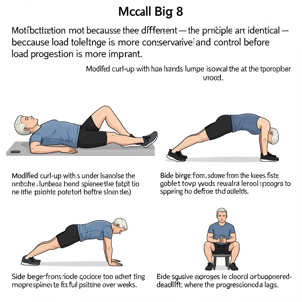
The McGill Big 3 are appropriate and important after 50, but they represent a starting point, not a ceiling. Once the Big 3 are performed with good technique and moderate comfort, progressive resistance exercise — squats, deadlifts, rows, and carries performed with conservative loads and excellent technique — are the most effective interventions for maintaining muscle mass, bone density, and spinal support capacity into older age. Consult a physiotherapist or exercise physiologist for a progression appropriate to your starting point.
The Social Isolation and Pain Amplification Pattern
Chronic pain in the over-50 population is frequently accompanied by progressive social withdrawal that amplifies pain through two mechanisms: first, physical activity reduces (removing its pain-modulating and mood-regulatory effects); second, social isolation itself activates the same neural pathways as physical threat and amplifies central pain sensitisation.
Retirement, the empty nest, bereavement, and the loss of the work-based social structure that many people rely on all converge in the 50-70 decade and create conditions for significant depression and pain escalation. Addressing this pattern requires recognising it — the person who "just has a bad back" is often also profoundly lonely and sedentary, and addressing only the mechanical component while ignoring the social one produces partial results at best.
In Review
- After 50, disc dehydration and height loss, facet OA, ligamentum flavum thickening, and declining bone density all progress — but these changes do not inevitably mean increasing pain.
- Late-stage disc degeneration often naturally stabilises and reduces pain as segments become self-limiting in their movement — this is the biological basis for the pain improvement many people experience in their 60s.
- Lumbar stenosis emerges from the combination of disc height loss, ligamentum flavum thickening, and facet OA; its hallmark is neurogenic claudication relieved by forward bending and aggravated by extension.
- Extension-biased rehabilitation is appropriate for disc herniations but contraindicated in significant lumbar stenosis — assessment is essential before choosing an exercise direction.
- Vertebral fragility fractures can occur with trivial loading in osteoporotic spines; weight-bearing and resistance exercise are the most effective interventions for bone density maintenance.
- Sarcopenia reduces paraspinal muscle mass by approximately 1% per year without resistance training, progressively increasing spinal load on passive structures.
- The McGill Big 3 remain appropriate and important after 50, with conservative load progression; resistance training beyond the Big 3 is needed to address sarcopenia.
- Social isolation in this decade amplifies pain sensitisation and must be addressed alongside the mechanical components of back pain management.