Osteoporosis and the Spine: Fracture Risk and Safe Exercise
How bone density loss affects spinal integrity, which activities carry real fracture risk, and the exercise prescription that builds bone without endangering it.
Bone as Living Tissue
Bone is not static scaffolding. It is metabolically active tissue that is continuously remodeled in response to mechanical loading, hormonal signals, nutrition, and age. Osteoblasts lay down new bone matrix; osteoclasts resorb old bone. In youth, this process favors formation. Peak bone mass is typically reached in the third decade of life. After that, the balance gradually shifts toward resorption.
Osteoporosis is the clinical expression of this imbalance taken to a level where bone microarchitecture is compromised and fracture risk becomes clinically significant. The World Health Organization defines osteoporosis as a bone mineral density (BMD) T-score at or below -2.5, measured by dual-energy X-ray absorptiometry (DEXA) scanning. Osteopenia — reduced bone density that has not yet reached the osteoporotic threshold — is defined as a T-score between -1.0 and -2.5.
These numbers matter enormously for how exercise is approached. Someone with a T-score of -1.5 and no prior fractures can exercise quite aggressively. Someone with a T-score of -3.0 and a previous vertebral compression fracture requires a substantially modified approach. The exercise prescription is not the same for everyone with "low bone density," and generalizing from one category to the other can cause harm.
How the Spine Is Affected by Osteoporosis
The vertebral bodies are composed largely of trabecular (cancellous) bone — the spongy inner framework of interconnecting struts. Trabecular bone has a much higher surface area than cortical (dense outer) bone and is therefore far more metabolically active and more rapidly affected by the hormonal and nutritional changes associated with osteoporosis. The lumbar and thoracic vertebrae are predominantly trabecular in structure, which is why these are the sites where osteoporotic fractures occur first and most commonly.
A vertebral compression fracture occurs when the trabecular bone of a vertebral body is no longer able to support normal compressive load. In severe osteoporosis, fractures can occur during activities that most people would consider low-risk: coughing, bending forward to retrieve an object from the floor, or reaching overhead. In milder osteoporosis, fractures typically require a meaningful loading event, but the threshold is substantially lower than in normal bone.
Vertebral compression fractures occur almost exclusively in the anterior vertebral body. The typical mechanism is forward flexion with or without added load. The anterior column bears the majority of compressive force during forward bending, and the trabecular bone in the anterior vertebral body fails under that compression. This is why the pattern of thoracic kyphosis that develops in osteoporotic patients — multiple anterior wedge fractures at adjacent levels — is so clinically distinctive.
The most important movement restriction for people with osteoporosis is forward spinal flexion under load. This means avoiding exercises that involve bending forward while carrying weight, trunk flexion against resistance, and high-repetition tasks involving reaching forward and down. Sit-ups, crunches, and exercises that curl the trunk forward are specifically contraindicated in moderate-to-severe osteoporosis.
DEXA Scanning and Risk Assessment
DEXA scanning is the gold standard for bone density measurement and provides T-scores and Z-scores for the lumbar spine and proximal femur. The T-score compares your bone density to a young healthy adult peak; the Z-score compares it to age-matched peers. For clinical fracture risk assessment, the T-score is the relevant figure.
DEXA scanning alone, however, does not fully capture fracture risk. The FRAX tool (Fracture Risk Assessment Tool), developed by the WHO, combines BMD with clinical risk factors — age, sex, prior fractures, family history, smoking, alcohol use, glucocorticoid use, rheumatoid arthritis, and secondary causes of osteoporosis — to calculate a 10-year probability of hip fracture and major osteoporotic fracture. A person with a borderline T-score and multiple clinical risk factors may have higher fracture risk than someone with a lower T-score and no risk factors.
Vertebral end-plate fractures deserve special mention. These are distinct from complete compression fractures and involve fracture of the thin cortical plate that separates the vertebral body from the disc. They are extremely common on MRI in older adults and are frequently asymptomatic. However, end-plate fractures allow the nucleus pulposus to intrude into the vertebral body (Schmorl's nodes) and alter the mechanical behavior of the affected segment. They are also markers of overall skeletal fragility.
DEXA scanning is recommended for all women over 65, men over 70, postmenopausal women under 65 with additional risk factors, and any adult who has experienced a low-trauma fracture after age 50. Many people with clinically significant bone density reduction have never been scanned — if you are in one of these categories and have not had a DEXA scan, this is worth discussing with your physician.
Which Exercises Build Bone
The mechanical principle governing bone formation is Wolff's Law: bone remodels in response to the mechanical forces applied to it. Specifically, bone formation is stimulated by dynamic loading — loading that creates deformation of the bone tissue — rather than static or sustained loading. The loading must be above a certain threshold (habitual activities that the bone is already accustomed to do not stimulate new formation) and should be applied in varied directions to stimulate bone across multiple planes.
This has direct implications for exercise prescription:
Impact loading — walking, jogging, stair climbing, and impact activities — stimulates bone formation at the hip and lumbar spine. The impact must be meaningful; swimming, while excellent for cardiovascular health, does not load the skeleton and does not build bone. Cycling similarly provides minimal bone stimulus. Walking is the minimum impact activity with demonstrated osteogenic effect; jogging and higher-impact activities stimulate more bone formation but require that the skeleton already have sufficient integrity to tolerate the impact.
Resistance training — particularly loaded lifting exercises — provides high-magnitude bone loading in the directions specific to the exercise performed. A weighted squat loads the lumbar vertebrae, femoral neck, and proximal tibia simultaneously. Deadlifts load the lumbar spine under high compressive force. Hip abductor and extensor exercises load the femoral neck in the region most at risk for hip fracture. The evidence consistently shows that resistance training with progressive loading is among the most powerful osteogenic interventions available.
High-force muscular contractions, even without external load, create meaningful bone deformation at the muscle attachment sites. Jumping exercises generate ground reaction forces of three to seven times body weight, stimulating bone across the lower extremity and lumbar spine.
What to Avoid
For people with established osteoporosis (T-score below -2.5) or a prior vertebral fracture, several activity categories carry disproportionate fracture risk:
Forward trunk flexion under load is the primary contraindication. This includes exercises that involve bending the trunk forward against resistance, weighted sit-ups or crunches, bending forward to lift from the floor without maintaining a neutral spine, and activities that repetitively flex the trunk forward (repeated bending during cleaning, gardening, or manual labor).
High-impact activities without appropriate bone density. Running, jumping, and high-impact aerobics place compressive and impact loads on vertebrae that may be insufficient for these forces in moderate-to-severe osteoporosis. This does not mean all impact is contraindicated — modest impact walking is osteogenic even in osteoporosis — but the type of impact should be calibrated to the degree of bone loss.
Twisting under load — particularly rotational exercises like golf swings, racquet sports, and weighted trunk rotation exercises — creates torsional stress on the vertebral bodies that can produce burst-type fractures in severely osteoporotic spines.
Spinal flexion yoga poses — including seated forward bends, rounded-spine poses, and extreme hip flexion postures — create the same anterior compression mechanism as weighted forward bending and should be avoided or significantly modified.
A safe alternative to flexion-based spinal exercises for osteoporotic patients is spinal extension. Prone back extensions, supine thoracic extensions, and any movement that takes the spine into extension rather than flexion loads the posterior elements rather than the anterior vertebral bodies — the site of osteoporotic fracture. Gentle prone back extension is both safe and actively beneficial for thoracic kyphosis prevention.
Fall Prevention: The Often-Overlooked Priority
A fracture requires both susceptible bone and sufficient loading to cause failure. Reducing fracture risk therefore has two components: improving bone density and reducing the loads applied to bones. For the spine, this primarily means avoiding the positions and movements described above. For the hip — the site of the most clinically serious osteoporotic fractures — it primarily means preventing falls.
Hip fracture is associated with approximately 25–30% mortality in the year following injury and permanent functional decline in many survivors. The majority of hip fractures in older adults result from falls. Fall prevention is therefore as important as — and arguably more immediately impactful than — bone density improvement for reducing the catastrophic consequences of osteoporosis.
Exercise-based fall prevention has strong evidence. The most effective components are:
- Balance training: Single-leg standing, tandem walking, and perturbation exercises train the reactive postural responses that prevent falls
- Progressive strength training: Lower extremity and hip strength is directly associated with fall risk; weakness in hip abductors and knee extensors is among the strongest predictors of falls
- Gait retraining: Slowed gait speed and reduced stride length are modifiable risk factors for falls; walking programs and specific gait training improve both
- Tai chi: Consistently shown in randomized trials to reduce fall risk by improving balance, proprioception, and reactive control
Medication Context
Medications for osteoporosis — bisphosphonates (alendronate, risedronate, zoledronic acid), denosumab, and anabolic agents (teriparatide, romosozumab) — do not replace exercise but interact with it in important ways. Bisphosphonates reduce bone resorption and improve T-scores modestly but do not restore bone architecture; exercise still provides essential loading stimulus for maintaining and building bone quality. Teriparatide, a parathyroid hormone analog, directly stimulates bone formation and has the most dramatic effect on trabecular bone architecture — it creates bone that is more mechanically responsive to exercise loading.
For patients on bisphosphonates, the concern about atypical femoral fractures at high cumulative doses is real but the absolute risk is very low. These fractures are associated with specific radiographic prodromal signs and can be monitored. They do not constitute a reason to avoid exercise.
Building a Safe Exercise Program
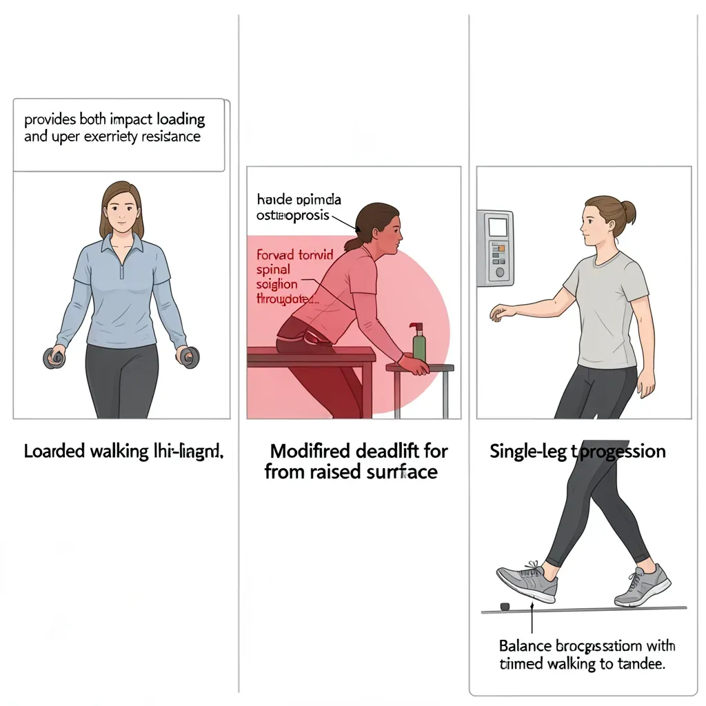
A practical exercise program for someone with osteopenia or osteoporosis should include the following elements:
Daily weight-bearing activity: At minimum, 30 minutes of brisk walking. For those with better bone density, progressing to intermittent jogging or step aerobics is appropriate.
Progressive resistance training: 2–3 sessions per week targeting the hip, spine, and upper extremity. Squats, modified deadlifts (from elevated surfaces in severe osteoporosis), hip abductor exercises, rows, and overhead pressing (with extension-biased spine position) are all appropriate.
Balance training: Incorporated into every session — single-leg stance, tandem standing, balance challenges during other exercises.
Extension-biased spine work: Prone back extensions, thoracic extension over a foam roller, and swimming backstroke are all loading spine in extension rather than flexion.
Avoid: Forward trunk flexion under load, high-impact twisting, sit-ups or crunches, heavy lifting from the floor with a flexed spine.
In Review
- Vertebral compression fractures occur in the anterior vertebral body under forward flexion loading; this is the primary movement restriction in moderate-to-severe osteoporosis
- DEXA T-scores classify bone density, but the FRAX tool combines clinical risk factors to provide a more accurate fracture risk estimate
- Bone formation is stimulated by dynamic, high-magnitude, varied-direction loading above the habitual threshold; swimming and cycling do not stimulate bone formation despite being beneficial for general health
- Resistance training with progressive loading and weight-bearing impact activities are the most osteogenic exercise interventions available
- Forward trunk flexion under load, high-impact rotational activities, and spinal flexion yoga poses carry specific fracture risk in osteoporotic spines and should be modified or avoided
- Fall prevention — through balance training, lower extremity strengthening, and gait training — is as important as bone density improvement for reducing hip fracture risk
- Medications for osteoporosis do not replace exercise; the exercise loading stimulus remains essential even in patients on bisphosphonates or anabolic agents
- Exercise for osteoporosis should be extension-biased for the spine, impact-appropriate for the hip, and progressively loaded within the limits of bone density and prior fracture history