The Paraspinal Muscles
The erector spinae, multifidus, and quadratus lumborum — what they do, how they fail, and how to rebuild them specifically.
Why Muscle Specificity Matters
Exercise programs for back pain are frequently designed as if muscles are interchangeable — as if any sufficiently hard "core workout" will produce the stability the injured spine needs. The evidence argues otherwise. Not all muscles contribute equally to spinal stability, not all muscles respond to injury in the same way, and not all muscles respond to the same training stimulus.
This chapter maps the muscles that matter most for lumbar spine stability, what each one actually does mechanically, how each one fails after injury, and what the evidence says about rebuilding each specifically. The Big 3 exercises introduced in Article 8 are not arbitrary — their selection is grounded in the anatomy and biomechanics described here.
The Two-System Model
Stuart McGill's foundational contribution to rehabilitation is the global-local distinction in trunk musculature.
The global muscle system consists of large, superficially positioned muscles that span multiple segments: the erector spinae group, rectus abdominis, and external obliques. These muscles produce gross movement — they flex, extend, and rotate the trunk. They generate high force but are not positioned to control the fine-grained motion of individual vertebrae. Stiffening the global system increases overall trunk stiffness but does not specifically prevent excessive motion at a single intervertebral joint.
The local muscle system consists of smaller, deeper muscles with direct vertebral attachments: multifidus, transversus abdominis, internal obliques, and the pelvic floor and diaphragm. These muscles cannot generate large forces, but they are positioned to monitor and control segmental motion directly. Their primary function is not to move the spine but to prevent specific patterns of intervertebral translation and rotation that would otherwise damage discs, facets, and ligaments.
A healthy spine relies on both systems working in coordination. After injury, the local system preferentially degrades — and because its failure is not visible in the way a large global muscle atrophy is, it frequently goes unaddressed.
Erector Spinae
The erector spinae group comprises three parallel columns running the length of the spine: iliocostalis (most lateral), longissimus (middle), and spinalis (most medial). In the lumbar region, they run from the sacrum and iliac crest upward, attaching to ribs, transverse processes, and spinous processes.
Their primary mechanical action is trunk extension — they are the primary spinal extensors in heavy lifting. During a deadlift, squat, or loaded forward bend, the erector spinae fire isometrically to prevent the spine from flexing under load. This is their most important stability role: eccentric and isometric control of trunk posture under load, not concentric trunk extension against resistance.
The erector spinae are essentially never weak in back pain patients in the clinical sense — they atrophy less dramatically than the local muscles and are adequately stimulated by most daily activities. However, they are frequently inappropriately over-recruited: patients who have lost local system control compensate by over-activating the erectors, producing a chronic, high-load, compressive pattern that fatigues the spine even during routine activity. The rehabilitation goal is not to strengthen the erectors further — it is to normalize their activation and allow the local system to share the load.
One common clinical observation: patients with chronic low back pain often have hyperactive, non-relaxing erector spinae. Instead of the normal pattern of cyclic activation (high during loading, low during relaxation), these muscles maintain sustained low-grade activation even at rest. This pattern is associated with elevated compressive spinal load throughout the day and poor sleep quality, because the muscles never fully unload. Learning to consciously relax the erectors during sedentary periods — not just strengthening them — is a legitimate rehabilitation target.
Multifidus
The multifidus is the most important muscle for understanding why specific rehabilitation works better than general exercise for spinal instability. It is a deep, segmentally organized muscle running from the sacrum to the cervical spine, with fascicles spanning one to four vertebral levels. Crucially, each fascicle has a direct attachment to the lamina and spinous process of the vertebra at its origin.
This architecture is unique: no other trunk muscle has direct segmental vertebral attachments at every level. This makes the multifidus a dedicated segmental stabilizer — when a specific intervertebral joint is loaded, the multifidus fascicle at that level is precisely positioned to resist unwanted translation and rotation.
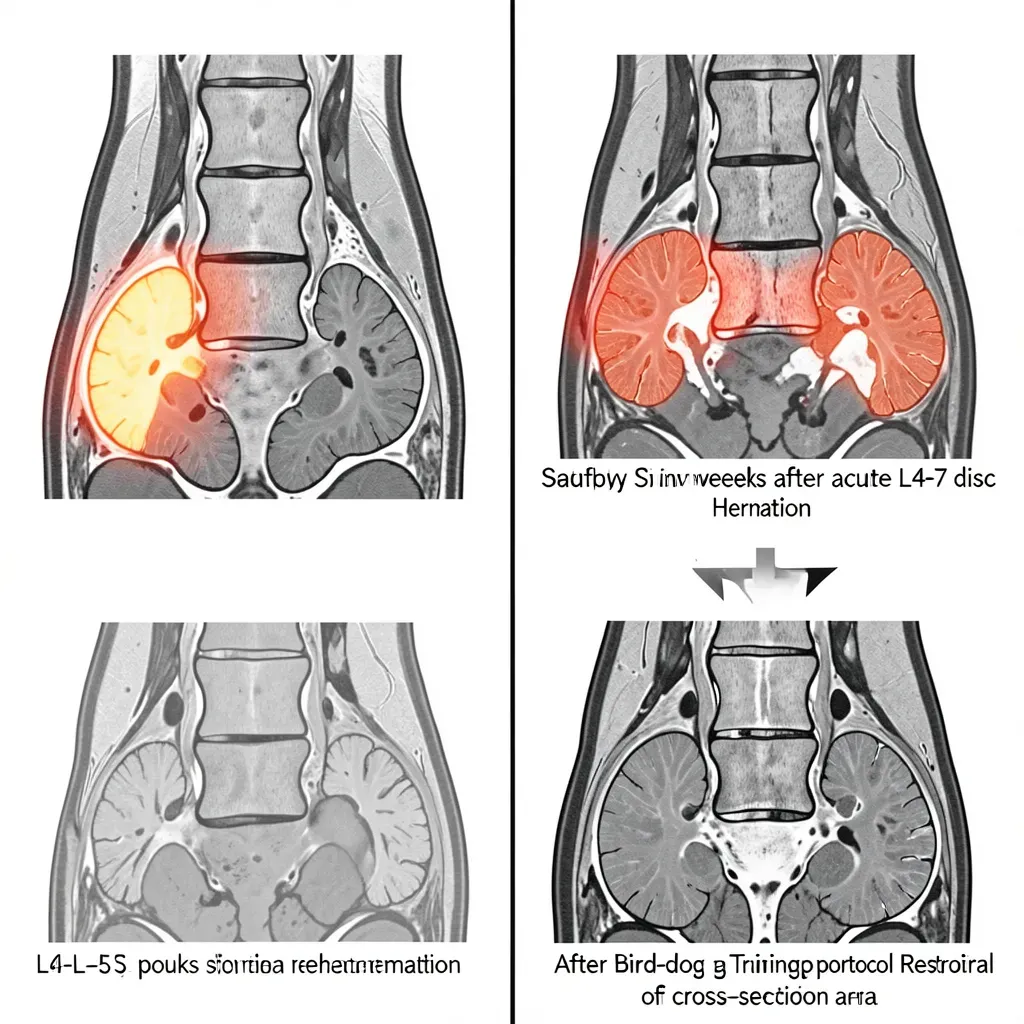
The atrophy problem: Multiple imaging studies using cross-sectional MRI have documented that multifidus atrophy occurs rapidly after acute low back pain — within weeks of injury onset — and is specific to the injured level. An L4-L5 disc herniation produces significant multifidus atrophy at L4-L5 but not at adjacent levels. This specificity confirms that the atrophy is neurologically mediated (inhibition of the specific segmental motor neurons), not simply global deconditioning.
The non-recovery problem: The critical finding from Hides, Richardson, and Jull (1996) is that multifidus atrophy does not spontaneously recover after resolution of the acute pain episode. Patients who recovered symptomatically but did not receive specific multifidus rehabilitation showed persistent atrophy at 10-week follow-up. This is the biological basis for the extremely high recurrence rate of low back pain — the underlying segmental instability from multifidus atrophy is never addressed, and the spine remains vulnerable to re-injury.
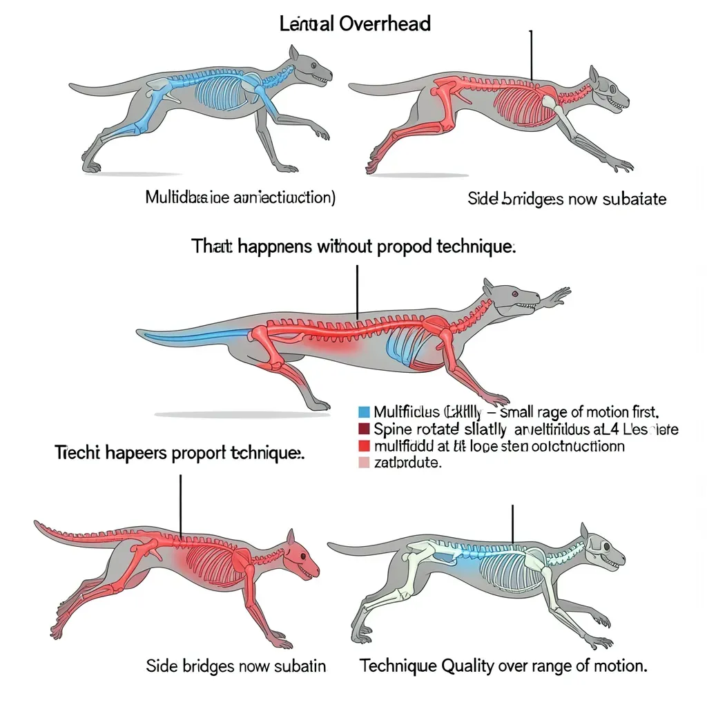
The rehabilitation implication: General exercise (walking, swimming, gym work) does not consistently restore the multifidus. The evidence supports specific low-load isometric activation of the multifidus, which is exactly what the bird-dog exercise achieves when performed correctly — sustained isometric contraction of the multifidus while the limbs are moving, with zero spinal motion.
Ultrasound imaging studies show that the multifidus activates best when the spine is held in a neutral position under a modest challenge. It is significantly less active during high-load exercises performed with spinal movement. This is why high-intensity back extensions on a Roman chair — a popular "lower back exercise" — do not effectively train the multifidus despite the high load. The multifidus is not a spinal mover; it is a spinal stabilizer. Training it requires sustained neutral position under a manageable challenge, not high-range-of-motion loading.
Quadratus Lumborum
The quadratus lumborum (QL) is a quadrilateral muscle in the posterior abdominal wall, running from the posterior iliac crest to the 12th rib and the transverse processes of L1-L4. It crosses the lumbar spine laterally and functions primarily as a lateral trunk stabilizer — resisting lateral flexion and providing frontal-plane stability to the lumbar spine.
The QL is also the critical muscle for hip-spine coupling in the frontal plane. During single-leg stance — walking, stair climbing, any unilateral activity — the body must resist a lateral tilt toward the unsupported leg. The QL on the stance side works in concert with the hip abductors to maintain pelvis-spine alignment. When either the QL or the hip abductors are weak, the pelvis drops and the lumbar spine laterally flexes to the unsupported side, compressing the facets and discs on the opposite side asymmetrically.
The side bridge as QL rehabilitation: The side bridge (side plank) in McGill's Big 3 is primarily a QL endurance exercise. Its value is in training sustained isometric lateral stability — the QL's actual function during walking and daily activity — rather than producing maximal force output.
Psoas Major
The psoas is the most controversial muscle in back pain rehabilitation, with strong opinions on multiple sides. The anatomy is not controversial: psoas arises from the lateral aspect of the lumbar vertebral bodies and transverse processes (T12-L5), travels anteriorly over the pelvic brim, and inserts on the lesser trochanter of the femur. When contracting with the femur fixed, it flexes the lumbar spine. When contracting with the spine fixed, it flexes the hip.
The debate concerns its stability role. Biomechanical models by Bogduk and Penning suggest the psoas line of action generates compressive load on the lumbar spine during hip flexion rather than reducing it — meaning psoas activation may not stabilize the spine but actually loads it. McGill's position is that psoas is primarily a hip flexor and that its contribution to lumbar stability is modest and variable.
The practical implication for rehabilitation: aggressive psoas stretching (often prescribed for anterior pelvic tilt) may or may not address the underlying problem, and psoas "strengthening" via loaded hip flexion exercises with an unstable spine (leg raises, certain crunch variations) increases lumbar compressive load without providing net stability benefit. For most patients, addressing the hip flexor-glute imbalance through hip extension training is more productive than psoas-specific work.
Gluteal Muscles as Indirect Stabilizers
The gluteus maximus and medius do not attach directly to lumbar vertebrae, but they profoundly influence lumbar loading through the pelvis. The glutes control anterior pelvic tilt (gluteus maximus extends the hip, reducing tilt) and lateral pelvic stability (gluteus medius controls frontal-plane hip position during single-leg stance).
In the absence of adequate gluteal activation, the lumbar spine compensates:
- For hip extension weakness: lumbar hyperextension substitutes for hip extension, loading the facets.
- For gluteus medius weakness: Trendelenburg gait pattern produces asymmetric lumbar compression.
The hip hinge pattern — the fundamental motor pattern behind safe lifting — is fundamentally a glute-dominant movement. Teaching the hip hinge correctly (hips back, spine neutral, loading through the glutes and hamstrings rather than through the lumbar extensors) is one of the highest-value interventions in back pain rehabilitation.
The endurance, not maximum strength, of the lumbar muscles is the strongest predictor of back pain risk. Biering-Sorensen's original 1984 study found that back extensor endurance — measured by the time a person can hold an unsupported horizontal trunk position — was the best predictor of future first-episode low back pain. Subsequent research by McGill confirmed that the endurance ratios among the muscles (extensor:flexor, right side bridge:left side bridge) matter as much as absolute values. Assess endurance capacity, not just strength, and train accordingly.
In Review
- The global muscle system produces trunk movement; the local system controls individual vertebral segments — only the local system can prevent the specific patterns of intervertebral motion that damage discs and facets.
- Multifidus atrophy after low back pain is rapid, level-specific, and neurologically mediated; it does not spontaneously recover with pain resolution and requires specific rehabilitation.
- General exercise is not equivalent to specific multifidus training; the bird-dog works because it trains sustained isometric activation in a neutral spine, not because it produces maximal load.
- The quadratus lumborum is the primary lateral stabilizer; the side bridge trains its endurance function, which is what actually matters for daily activity.
- The psoas primarily flexes the hip and loads the lumbar spine under contraction — treat it as a hip flexor, not a spinal stabilizer.
- Gluteal weakness causes lumbar compensation during hip extension and single-leg stance, directly increasing facet and disc load; hip hinge training is among the most important rehabilitation movements.
- Lumbar muscle endurance, not maximal strength, is the strongest predictor of back pain risk; endurance ratios between muscle groups matter as much as absolute values.