The Hip-Spine Relationship
How hip mobility, hip strength, and pelvic position directly determine lumbar spine loading — and why many back problems originate at the hip.
Two Joints, One Movement System
The lumbar spine and the hip do not operate independently. Every movement that involves the lower extremity creates a mechanical demand on the lumbar spine unless the hip is functioning well — mobile enough to absorb range of motion, strong enough to absorb force, and positioned correctly in the pelvis. When the hip fails on any of these fronts, the lumbar spine compensates.
This compensation is not always dramatic. It can be as subtle as a few extra degrees of lumbar flexion at the bottom of a forward bend, or a slight rotation of the pelvis during single-leg stance. But repeated thousands of times per day, accumulated over years, those small compensations become the primary mechanical driver of disc, facet, and ligament pathology.
The clinical implication is significant: a patient may have genuine lumbar disc pathology — confirmed on imaging — whose primary cause is a hip mobility or strength deficit that predates the disc injury by years. Treating the disc without addressing the hip problem is treating the symptom without treating the cause.
Lumbopelvic Rhythm
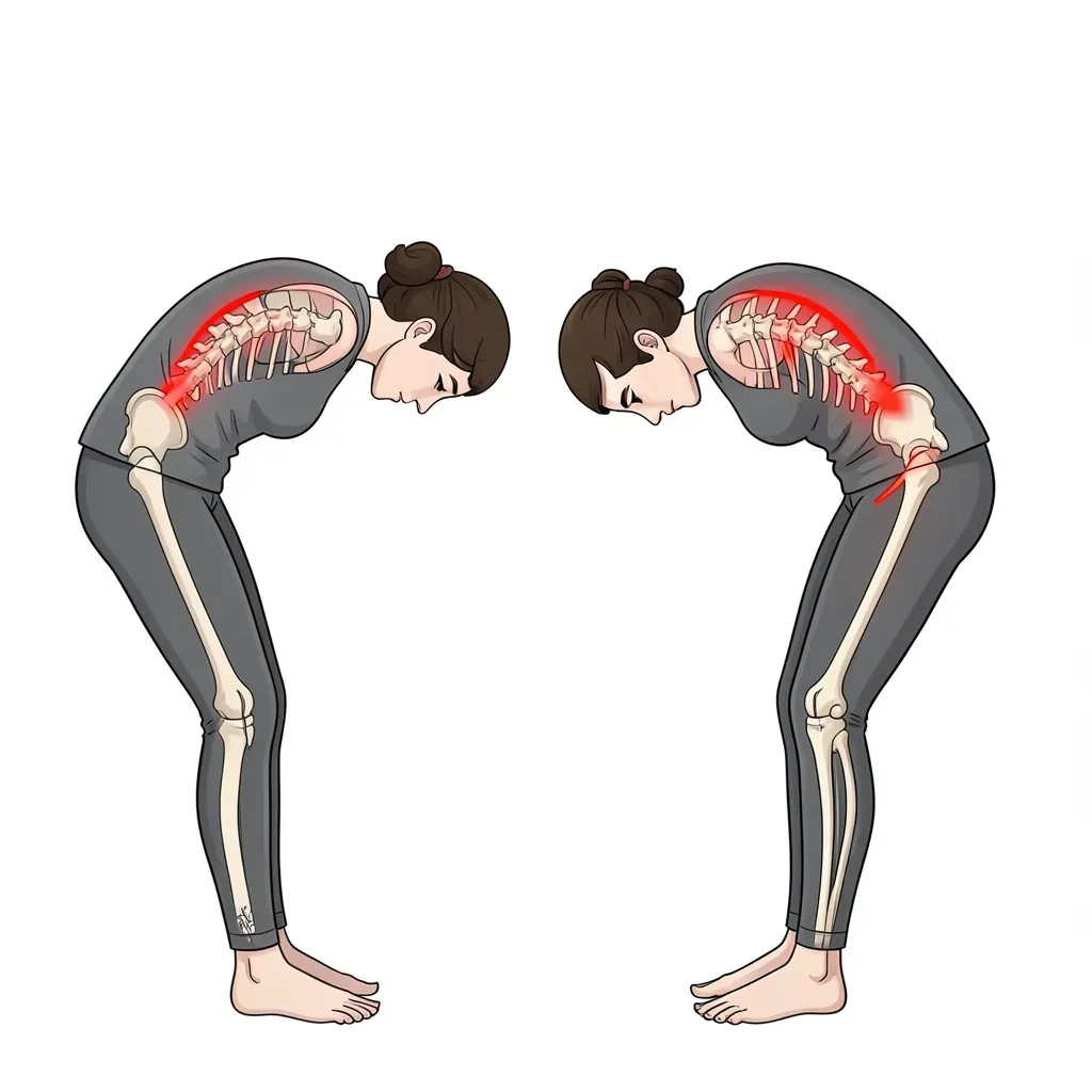
Lumbopelvic rhythm describes the coordinated movement of the pelvis and lumbar spine during forward bending. In a well-functioning system, forward bending involves two simultaneous contributions: lumbar flexion (flattening of the lumbar curve) and pelvic anterior rotation (hip flexion). The ratio of lumbar flexion to pelvic rotation varies during the movement — early bending is dominated by lumbar contribution; continued bending is dominated by hip contribution as the hamstrings allow the pelvis to rotate forward.
When hip range of motion is limited — tight hip flexors, limited hip internal rotation, or femoroacetabular impingement (see below) — the pelvis cannot contribute its normal share of the range of motion during forward bending. The lumbar spine must compensate by moving further into flexion than it otherwise would. This is called "lumbar-dominant" lumbopelvic rhythm, and it concentrates strain on the posterior disc annulus and posterior spinal ligaments during every forward bending task.
The same compensation occurs in reverse during rising: without adequate hip extension range, the pelvis tips anteriorly to allow the body to extend, and the lumbar spine hyperextends compensatorily. This produces facet loading every time the patient rises from a chair, steps up a stair, or walks.
The forward bend test is one of the most revealing assessments in spine rehabilitation. Stand with feet shoulder-width apart and bend forward, trying to touch the toes. A therapist watching from the side can observe the segmental contributions: does the lumbar curve flatten smoothly, or does it remain lordotic (indicating lumbar extension dominance)? Does the pelvis rotate forward freely, or does it stop early while the lumbar spine accelerates its flexion? The test localizes the stiffness — the joint that stops moving earliest is the joint creating the compensation elsewhere.
Hip Internal Rotation and Compensatory Lumbar Rotation
Hip internal rotation — the ability to rotate the femur inward within the acetabulum — is one of the most underappreciated mobility deficits in back pain. The normal range is approximately 30-45 degrees in adults, and it declines substantially with age, sedentary behavior, and hip osteoarthritis.
The lumbar spine is not designed to tolerate axial rotation. The annular fiber architecture of the disc resists rotation specifically, and the facet joint orientation in the lumbar spine limits rotation to approximately 3-5 degrees per level before capsular restraint is engaged. When the hip cannot provide adequate internal rotation during a walking, squatting, or stepping movement, the pelvis and lumbar spine rotate to compensate.
This rotation is small per step — perhaps one or two degrees — but the cumulative effect across thousands of daily steps is significant. McGill's work has demonstrated that cumulative rotation is among the most damaging loading patterns for the lumbar annulus, producing delamination of the annular lamellae through fatigue failure. Hip internal rotation limitation is therefore a direct upstream cause of lumbar disc annular injury.
Clinical test: Seated hip internal rotation measurement. Sit at the edge of a table with hips and knees at 90 degrees. Rotate the foot outward (which creates internal rotation at the hip) as far as possible without moving the pelvis. Normal is 30-45 degrees. Less than 25 degrees, especially if asymmetric, is clinically significant for lumbopelvic mechanics.
Femoroacetabular Impingement and Its Effect on Lumbar Loading
Femoroacetabular impingement (FAI) is a morphological condition in which abnormal bony contact between the femoral head (cam-type FAI) or acetabular rim (pincer-type FAI) limits hip range of motion — particularly hip flexion combined with internal rotation.
FAI is common and frequently underdiagnosed. Population studies suggest cam morphology is present in 20-40% of adults and is more common in athletes who participated in sport during skeletal development. The key clinical relevance for back pain is this: when hip flexion is blocked by impingement, forward bending requires additional lumbar flexion to compensate. Deep squat positions require lumbar flexion to achieve the depth otherwise provided by hip flexion. Any activity requiring combined hip flexion and internal rotation — picking up objects from the ground, squatting, cycling — increases lumbar load when FAI limits hip range.
Patients with undiagnosed FAI often present with low back pain as the primary complaint because the spine is absorbing the movement the hip cannot provide. A groin crease pinch during hip flexion, pain with combined hip flexion-adduction-internal rotation (FADIR test), or a pattern of low back pain that is consistently worse with hip-dominant activities should prompt evaluation for hip impingement.
FAI is often treated surgically with hip arthroscopy, but surgery addresses the bony morphology and labral damage — not the learned compensation patterns. Patients who have FAI-related surgery frequently need specific rehabilitation to retrain the hip to move through its newly available range and to undo the compensatory lumbar motion patterns developed over years. Back pain that is actually FAI-driven does not reliably improve after spine surgery — the hip morphology is still limiting range of motion and the spine is still compensating.
Anterior Pelvic Tilt Mechanics
Anterior pelvic tilt — the pelvis tilting forward such that the anterior superior iliac spine moves forward and down relative to the posterior superior iliac spine — increases lumbar lordosis. The practical consequence is increased facet joint compression, reduced disc posterior margin opening, and increased tension on the posterior hip capsule.
The common narrative that anterior pelvic tilt is always pathological is not accurate — some degree of anterior tilt is normal and appropriate. The problem arises when the tilt is excessive or habitual, and when the person cannot voluntarily reduce it during loading.
The primary drivers of excessive anterior tilt are:
- Hip flexor tightness (particularly the iliopsoas and rectus femoris), which pulls the pelvis anteriorly from the front
- Gluteal weakness, which reduces the posterior pulling force on the pelvis from behind
- Hamstring laxity (paradoxically — loose hamstrings allow more anterior tilt without the posterior check they normally provide)
Correcting the tilt involves both passive tissue work (hip flexor stretching, specifically the rectus femoris and iliopsoas in full hip extension) and active motor retraining (teaching the patient to posteriorly tilt the pelvis using the glutes in functional positions — standing, squatting, hinging).
The Tight Hip Flexor — Weak Glute Pattern
This is the most clinically common hip-spine dysfunction pattern. Prolonged sitting shortens the hip flexors adaptively while simultaneously inhibiting the gluteals through reciprocal inhibition (when a muscle is in a shortened position for prolonged periods, its antagonist is neurologically inhibited). The resulting pattern is:
- Shortened, stiff hip flexors that resist full hip extension
- Inhibited, poorly activating gluteals that cannot generate adequate hip extension force
- Compensatory lumbar hyperextension during any activity requiring hip extension — walking, stair climbing, standing from a chair, gym exercises
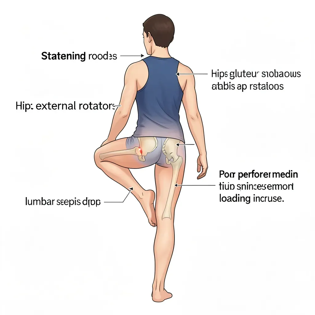
The hip airplane exercise (single-leg stance with controlled trunk rotation using hip external rotators) and the hip 90/90 stretch (targeting both internal and external rotation range of motion) are targeted interventions for this pattern, which is why they appear repeatedly in the mobility component of this program.
Testing Hip Mobility
Three assessments provide the most clinically useful information:
Thomas test (hip flexor length): Lying supine, pull one knee to the chest and observe the contralateral leg. If the contralateral thigh rises off the table, the hip flexors (primarily iliopsoas) are shortened on that side. If the knee extends (moves toward straight), the rectus femoris is the tighter component.
Seated hip internal rotation (described above): Less than 25 degrees is significant; asymmetry greater than 10 degrees between sides is significant.
Hip extension in prone (Ely's test variant): Lying prone, passively flex the knee and bring the heel toward the buttock. Note whether the ipsilateral hip rises off the table (rectus femoris restriction) and how much range is available before the lumbar spine begins to extend.
These three tests, performed together, identify the primary tissue contributors to hip-spine dysfunction and guide which interventions are priorities.
Asymmetric findings on hip mobility tests are more clinically meaningful than absolute bilateral restriction. If your left hip internal rotation is 40 degrees and your right is 15 degrees, that asymmetry directly translates into asymmetric lumbar rotation compensation during every left-step of your gait cycle. The most important correction is always to the side with the greater restriction, regardless of whether the other side is in the normal range.
In Review
- The hip and lumbar spine are mechanically coupled — hip mobility and strength deficits are directly compensated by increased lumbar motion and loading.
- Lumbopelvic rhythm during forward bending requires adequate hip flexion range; hip restriction produces lumbar-dominant bending that concentrates strain on the posterior disc and ligaments.
- Hip internal rotation loss of less than 25 degrees or greater than 10 degrees of side-to-side asymmetry produces compensatory lumbar rotation that accumulates as annular fatigue damage over time.
- FAI limits hip flexion combined with internal rotation; patients with FAI frequently present with back pain as the primary complaint because the spine compensates for what the hip cannot provide.
- Anterior pelvic tilt is driven by hip flexor shortening and gluteal weakness and produces chronic facet loading; correction requires both tissue work and motor retraining.
- The tight hip flexor — weak glute pattern is the most common hip-spine dysfunction, caused by prolonged sitting and directly testable with the Thomas test.
- Asymmetric hip mobility findings are more clinically significant than bilateral restriction; always prioritize the more restricted side.