Disc Nutrition & Health
How intervertebral discs get nutrients without a blood supply, why they degenerate, and what you can do to slow the process.
The Disc Is Not a Passive Spacer
Intervertebral discs are often described in lay terms as "shock absorbers" or "cushions," implying they are passive structural elements that either hold up or fail. This framing misses almost everything important. Discs are living tissue — their cells actively maintain the extracellular matrix, respond to mechanical signals, regulate hydration, and either preserve or degrade disc architecture depending on the metabolic environment they operate in.
Understanding disc nutrition is not an academic exercise. Every major controllable risk factor for disc degeneration — smoking, sedentary behavior, obesity, poor sleep — operates partly through the disc nutrition pathway. Understanding the mechanism gives you a coherent model for why the lifestyle interventions in this program work, and why some people's discs age faster than their chronological age would predict.
The Avascular Problem
Most tissues in the body receive oxygen and glucose directly from the capillary network embedded in them. Discs cannot do this. After the first decade of life, the blood vessels that originally supplied the disc interior regress, and the disc becomes one of the largest avascular structures in the human body.
Disc cells — chondrocyte-like cells embedded in the nucleus and annulus — must obtain all nutrients by diffusion. Oxygen and glucose diffuse inward from the disc periphery and through the cartilaginous endplates from the vertebral body vasculature. Metabolic waste products (primarily lactate) diffuse outward along the same concentration gradients.
The distances involved are significant. The center of the nucleus pulposus in an adult lumbar disc can be 8mm or more from the nearest blood supply. At this distance, oxygen tension at the nucleus center is approximately 1-5% — barely above the anaerobic threshold. The disc lives in a chronically hypoxic state and is metabolically fragile as a result.
The Endplate as the Gateway
The cartilaginous endplates are the primary route for disc nutrition. They are thin (approximately 0.6mm) plates of hyaline cartilage covering the superior and inferior disc surfaces, embedded with a fine network of nutrient channels called vascular buds that originate in the underlying vertebral bone.
Endplate integrity is the single most important determinant of disc nutrition. Damage to the endplates — from acute compressive overload, Schmorl's nodes (vertical disc herniations through the endplate), or the progressive calcification that accompanies disc degeneration — reduces nutrient transport and accelerates the degeneration cascade.
Vertebral endplate signal changes on MRI (Modic changes) are among the strongest imaging predictors of chronic low back pain — far more predictive than disc height loss or disc bulge alone. Modic Type 1 changes (bone marrow edema adjacent to the endplate) indicate active endplate inflammation and are associated with particularly severe pain presentations.
Modic changes, when present on MRI, are worth understanding. Type 1 (hypointense on T1, hyperintense on T2) indicates active inflammation — often associated with pain that is severe and constant. Type 2 (hyperintense on T1) indicates fatty replacement of marrow — often associated with more stable, less acute symptoms. Type 3 (hypointense on both) indicates sclerosis. The transition from Type 1 to Type 2 over time generally correlates with symptom improvement. This timeline is measured in months to years, not weeks.
The Pump Mechanism
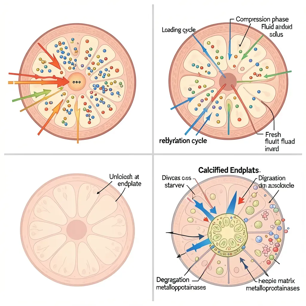
Because diffusion through the avascular disc is slow, the disc relies on a mechanical assist: the pressure changes generated by movement act as a pump, transiently deforming the disc and driving fluid and solute exchange across the endplate.
Loading compresses the nucleus, increasing hydrostatic pressure and driving fluid outward. Unloading (supine rest, standing decompression) allows the disc to rehydrate and draws fresh nutrient-rich fluid inward. This cyclical loading-unloading is not just tolerated by the disc — it is required for adequate nutrition.
This is why the optimal spinal health prescription is not rest, but varied movement. Sustained postures — prolonged sitting, prolonged standing in one position, sustained bed rest — reduce the pumping action and create a nutritional stagnation in the disc center. Exercise, walking, and positional changes throughout the day maintain the pump mechanism.
The disc is approximately 1-2mm taller in the morning after overnight horizontal rest (maximum rehydration) and decreases in height by approximately the same amount over the course of the day's loading. This diurnal variation in disc height is normal and expected. The clinical implication: spinal loads are highest and disc stiffness greatest in the morning, which is why McGill recommends avoiding heavy lifting and end-range bending in the first 30-60 minutes after rising.
Spinal flexion exercises — toe touches, sit-ups, spine stretches — are most dangerous in the morning when the disc is maximally hydrated and internal pressure is highest. The annular fibers under a well-hydrated nucleus are under greater tension and are more susceptible to tearing during flexion. This is not an argument against movement in the morning; it is an argument against aggressive end-range spinal flexion before the disc has partially dehydrated and adjusted to the day's loading pattern. Walking for 15-20 minutes first is a simple and effective way to begin that adjustment.
The Degeneration Cascade
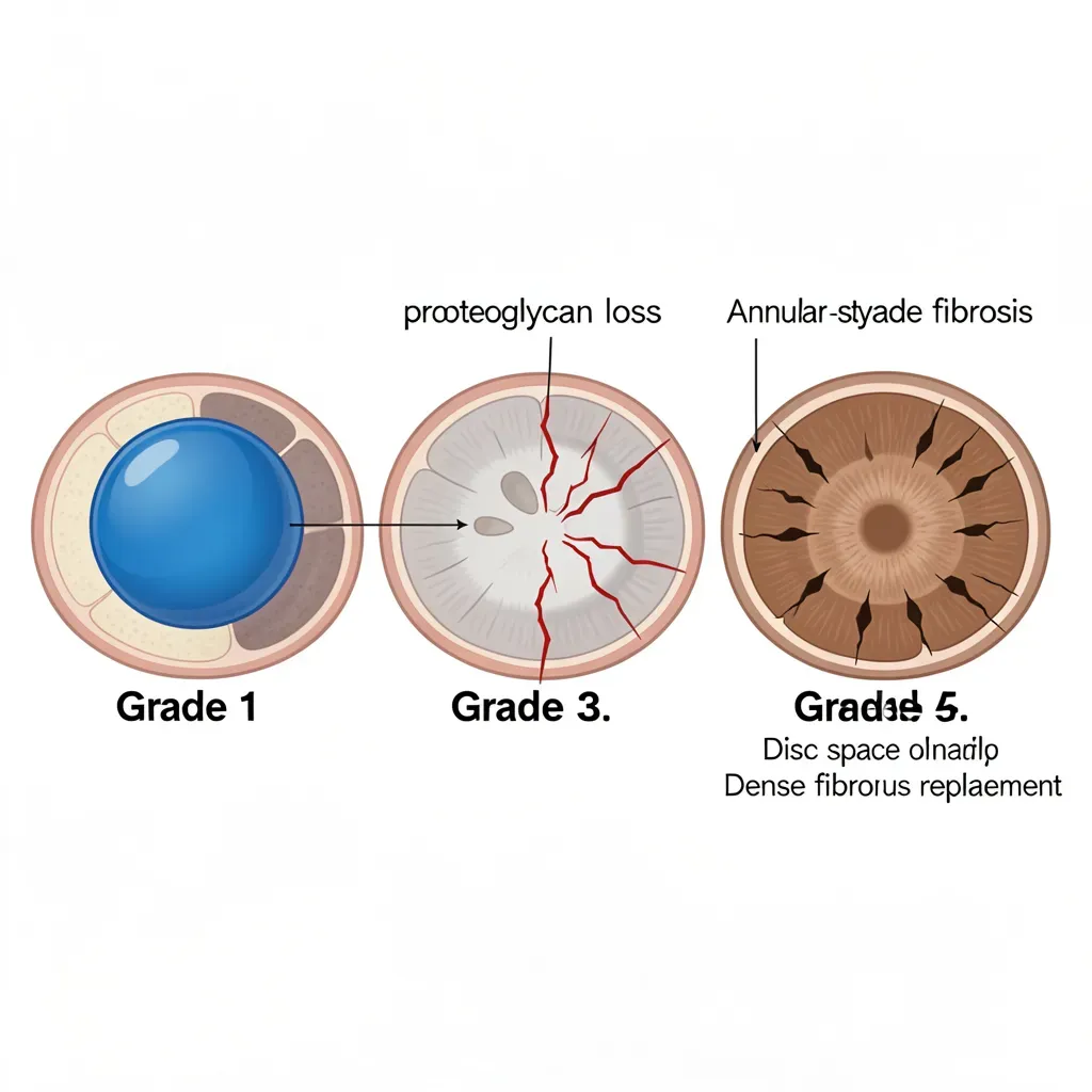
Disc degeneration is not a single event — it is a cascade. Thompson et al. described five grades of disc degeneration visible on gross pathology specimens, and the cascade follows a predictable biological sequence.
Grade 1-2 (Normal to Early Change): The nucleus is gelatinous and well-hydrated. The annular lamellae are discrete and intact. Minor endplate irregularities may be present. Pain is rare at this stage.
Grade 3 (Moderate Degeneration): Nuclear material becomes less gelatinous and more fibrous. The nucleus begins to desiccate — losing water-binding proteoglycans (primarily aggrecan). Disc height begins to decrease. Small radial tears may appear in the annulus. This is the stage where pain becomes possible, particularly from annular fissures that penetrate the outer third of the annulus (which is innervated).
Grade 4-5 (Severe Degeneration): The nucleus is largely replaced by fibrous tissue and shows no hydraulic behavior. Disc height is significantly reduced. Diffuse annular disruption. End-stage degeneration paradoxically may produce less pain than moderate degeneration in some patients — because the sensitized, torn annular fibers have been replaced by relatively inert scar tissue.
The cascade is driven primarily by the failure of nucleus cells to maintain proteoglycan production. Without adequate proteoglycans, the nucleus loses its water-binding capacity, osmotic pressure drops, the hydraulic mechanism fails, and mechanical load is transferred to the annulus in ways it was not designed to bear.
Smoking and Disc Nutrition
Smoking is the most potent modifiable risk factor for accelerated disc degeneration, and the mechanism is direct: nicotine causes vasoconstriction of the small vessels feeding the vertebral endplates. Reduced blood flow to the subchondral bone reduces the nutrient concentration gradient across the endplate, decreasing diffusion rates into the disc center.
Multiple large epidemiological studies confirm the association. Smokers show disc degeneration approximately 10 years earlier than non-smokers when controlling for age, occupation, and physical activity. The effect is dose-dependent — pack-years of smoking history correlates directly with degeneration grade. The good news: cessation allows partial vascular recovery, and the acceleration appears to slow (though not fully reverse) after quitting.
Carbon monoxide from cigarette smoke adds a second mechanism: it competes with oxygen for hemoglobin binding, reducing overall tissue oxygenation. The disc, already barely above its anaerobic threshold, is particularly sensitive to any further reduction in oxygen delivery.
Genetics vs. Lifestyle
Twin studies — particularly the Finnish and Danish back pain twin registries — have established that approximately 60-70% of disc degeneration variance is attributable to genetic factors. The genes involved primarily regulate proteoglycan synthesis, collagen composition, and the inflammatory response within the disc. This finding is often misinterpreted as fatalistic: "my discs were always going to degenerate."
The correct interpretation is more nuanced. Genetic factors determine your baseline disc vulnerability — how quickly your discs would degenerate under average loading conditions. Lifestyle factors — smoking, sedentary behavior, obesity, heavy occupational loading without adequate recovery — determine how far above or below that baseline trajectory you actually fall. A person with poor disc genetics who remains active, maintains healthy body weight, never smokes, and protects their spine during loading may degenerate slower than a person with favorable genetics who smokes, sits for 10 hours daily, and carries 40 pounds of excess body weight.
"Disc degeneration" on an MRI report is almost universally present in adults over 40 and is present in more than 30% of adults by age 30 — in the complete absence of back pain. The imaging finding alone does not explain symptoms. What matters clinically is whether the specific degenerated segment is the source of nociceptive or radicular symptoms, and whether the surrounding tissues (annulus, endplate, ligaments, facet joints) are contributing to the pain picture. A thoughtful clinician interprets imaging in the context of your physical examination and symptom pattern — not in isolation.
In Review
- Discs are avascular after early childhood; all nutrients diffuse through cartilaginous endplates from adjacent vertebral bone vasculature — making endplate integrity the critical chokepoint for disc health.
- The pump mechanism — cyclical loading and unloading during movement — drives convective fluid exchange and is essential for disc nutrition; sustained postures impair this mechanism.
- Discs are most hydrated and most vulnerable to flexion injury in the morning; the first 30-60 minutes after rising warrant conservative movement choices.
- The degeneration cascade proceeds through five Thompson grades, driven by proteoglycan loss in the nucleus, annular fissuring, and eventual fibrosis.
- Smoking accelerates degeneration by 10 years on average through nicotine-mediated endplate vasoconstriction and carbon monoxide-mediated oxygen reduction.
- Genetics accounts for 60-70% of degeneration variance — but lifestyle determines whether you fall above or below that genetic trajectory.
- Disc degeneration on imaging is almost universal in adults over 40 and does not equal clinical back pain; interpretation requires correlation with symptoms and physical examination.