Referred Pain and Radiculopathy: Where Pain Goes and Why
Why back pain appears in the leg, hip, or foot — the anatomical basis of referral patterns, the difference between referred pain and radiculopathy, and how to interpret your own symptom distribution.
Why Pain Travels
One of the most confusing aspects of lumbar spine pathology is that the pain rarely stays where the problem is. A damaged L5-S1 disc does not simply hurt at L5-S1. It may produce a dull ache deep in the buttock, sharp shooting pain down the back of the thigh, burning in the calf, numbness in the foot, or some combination of all of these. Meanwhile, the lower back itself may feel entirely normal on a given day.
This phenomenon — pain experienced at a location distant from its source — is called referred pain, and understanding its mechanisms is essential for interpreting your symptoms accurately, having productive conversations with clinicians, and not concluding that you have ten separate problems when you have one.
There are two fundamentally different mechanisms that cause pain to travel from the spine into the limb: referred pain and radiculopathy. They look superficially similar — both can produce leg symptoms — but their mechanisms, clinical features, and implications for treatment are distinct.
Referred Pain: The Convergence Mechanism
Referred pain occurs because the brain makes a mistake. It is not a metaphor; it is a literal misattribution of location.
Sensory neurons from many different body regions converge on the same interneurons in the dorsal horn of the spinal cord. When a nociceptive signal from a spinal structure (disc, ligament, facet joint, muscle) arrives at a dorsal horn level, it activates second-order neurons that also receive input from distant tissues — the buttock, the thigh, the groin. The brain, which must decide where in the body this signal is coming from, sometimes gets it wrong. It assigns the pain to the region it most commonly associates with that set of neurons — often a peripheral structure rather than the deep spinal source.
This is the same mechanism that causes cardiac pain to radiate into the left arm: sensory neurons from the heart and from the arm converge on the same spinal segments (C8-T1), and the brain's best guess at the pain's origin is wrong.
Referred pain from lumbar structures tends to be:
- Deep, aching, or cramping in character
- Diffuse and poorly localized — you can gesture roughly at the area but not point to a spot
- Reproduced by loading or stressing the specific spinal structure causing it
- Not associated with clear neurological deficit (weakness, reflex loss, dermatomal numbness)
- Variable in distribution — it does not follow the precise nerve root maps shown in anatomy textbooks
The distinction between referred pain and radiculopathy matters clinically because they respond to different treatments. Referred pain from a facet joint responds well to facet joint injections or medial branch blocks. Referred pain from a disc may improve with specific directional exercises. Radiculopathy may require nerve-focused interventions. A clinician who conflates the two will choose the wrong target.
Sclerotome Referral: Disc and Facet Patterns
Referred pain patterns from spinal structures follow sclerotome maps rather than dermatome maps. Sclerotomes are the embryonic segments from which bone, disc, and deep connective tissue develop — they differ significantly from dermatomes, which map the skin's surface nerve supply.
Kellgren's classic experiments in the 1930s and 1940s demonstrated this by injecting hypertonic saline into specific spinal structures in healthy volunteers and recording where pain was felt. Disc stimulation at L1-L2, for example, produces pain in the groin and anterior thigh — not the dermatome territory of those nerve roots. L4-L5 disc irritation produces deep buttock and posterior thigh pain. L5-S1 irritation produces pain into the posterior thigh and calf, often mimicking sciatica.
Facet joints have their own referral patterns, mapped by similar injection studies. The L4-L5 facet refers to the buttock and lateral thigh. The L5-S1 facet refers to the buttock and posterior thigh, extending as far as the popliteal fossa. These patterns overlap considerably with L4-L5 and L5-S1 disc referral, which is part of why clinical differentiation requires systematic examination rather than symptom location alone.
Radiculopathy: The Nerve Root Mechanism
Radiculopathy is a different animal. It results from mechanical compression, chemical irritation, or inflammation of a spinal nerve root — typically where it exits the intervertebral foramen or within the lateral recess. The most common cause is a disc herniation compressing the nerve root, though bony narrowing (foraminal stenosis from osteophytes or hypertrophied ligamentum flavum) is common in older patients.
When a nerve root is compromised, the entire peripheral distribution of that nerve can be affected. This produces a characteristic syndrome: pain, altered sensation, and potentially motor weakness or reflex loss, all following the specific territory of the affected nerve root — the dermatome.
Radiculopathy differs from referred pain in several clinically important ways:
Quality of sensation: Radicular pain is often described as sharp, shooting, electric, or burning — the signature of direct nerve fiber activation. Referred pain is typically deeper and duller.
Distribution: Radiculopathy follows dermatomal maps with reasonable consistency; the pain, numbness, or paresthesia tracks down a fairly specific strip of skin corresponding to the nerve root. Referred pain is more diffuse.
Neurological signs: Radiculopathy can produce measurable deficits — reduced sensation in a dermatome, weakness in a myotome, loss of a deep tendon reflex. Referred pain typically does not produce these findings.
Straight leg raise: The straight leg raise (SLR) test stretches the sciatic nerve and its contributing nerve roots. A positive SLR — reproduction of leg pain below the knee at less than 70 degrees of hip flexion — has reasonable sensitivity for L4-S1 nerve root compromise and is specific enough that a positive test in the contralateral leg (crossed SLR) is highly specific for large disc herniation.
Radiculopathy does not require disc herniation. Foraminal stenosis from facet hypertrophy, osteophyte formation, or spondylolisthesis can compress a nerve root without any disc herniation being visible on MRI. Conversely, many disc herniations visible on MRI do not contact a nerve root and produce no radiculopathy — imaging alone is not sufficient to diagnose or rule out radiculopathy.
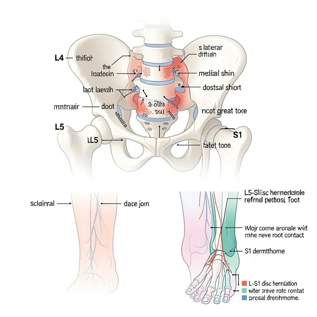
L4 Nerve Root: Anterior Thigh and Medial Leg
The L4 nerve root exits between L3 and L4 vertebrae. When compromised, it produces:
- Pain and paresthesia tracking from the lower lumbar region, across the lateral hip, down the anterior thigh, and into the medial shin and foot (the medial malleolus and great toe)
- Weakness in the quadriceps (knee extension) and hip flexors, which may manifest as difficulty climbing stairs or rising from a chair
- Diminished or absent patellar tendon reflex (the knee jerk)
- Positive femoral nerve stretch test: with the patient prone, passive knee flexion to 90 degrees followed by hip extension reproduces anterior thigh pain
L4 radiculopathy is less common than L5 or S1 because the L3-L4 disc is less frequently herniated than the lower levels, but it is clinically significant because the patellar reflex change is easily missed and weakness in the quad is often attributed to deconditioning.
L5 Nerve Root: Lateral Leg and Dorsal Foot
L5 is the most commonly compromised nerve root in disc herniation, given the frequency of L4-L5 pathology. Its distribution:
- Pain from the lateral lower back through the buttock, lateral thigh, lateral leg, and dorsum of the foot to the great toe (the L5 dermatome)
- Weakness in foot dorsiflexion (tibialis anterior), toe extension (extensor hallucis longus), and hip abduction (gluteus medius)
- The classic "foot drop" — inability to fully dorsiflex the foot when walking, producing a steppage gait — is L5 weakness
- No standard reflex is clearly associated with L5 alone, which makes the neurological examination more reliant on sensory and motor testing
The L5 dermatome extends to the webspace between the first and second toes — testing sensation there versus the lateral border of the foot (S1) is a useful clinical discriminator.
S1 Nerve Root: Posterior Leg and Lateral Foot
S1 radiculopathy most commonly results from L5-S1 disc herniation. Its distribution:
- Pain from the lower back, through the center of the buttock, down the posterior thigh and calf, to the lateral border of the foot and the lateral three toes
- Weakness in plantarflexion (calf raise) and toe flexion
- Diminished or absent Achilles tendon reflex (the ankle jerk) — one of the most reliable neurological signs in lumbar radiculopathy
- Single-leg heel raise weakness: the patient stands on the affected leg and attempts a full heel raise; subtle weakness shows as reduced height or increased wobble compared to the other side
S1 is the most frequently implicated root in lumbar radiculopathy overall, largely because L5-S1 is the highest-load disc in the lumbar spine.
The Piriformis Problem
The piriformis muscle sits deep in the buttock and has a peculiar anatomical relationship with the sciatic nerve. In about 15% of the population, the sciatic nerve (or one of its divisions) passes through the piriformis rather than beneath it. When the piriformis is hypertonic, inflamed, or enlarged, it can compress or irritate the sciatic nerve directly — producing a syndrome that mimics L5-S1 radiculopathy: buttock pain, posterior leg pain, and sometimes paresthesia into the foot.
This is clinically important because piriformis syndrome responds to different treatments than nerve root compression (stretching, soft tissue release, and occasionally injection versus spinal interventions), and because MRI of the lumbar spine will appear entirely normal, which can be dismissed as "no findings" without the correct diagnosis.
Differentiating features include: pain that is worsened by sitting (especially for prolonged periods), tenderness to deep palpation of the buttock, and positive FAIR test (hip Flexion, Adduction, and Internal Rotation reproducing sciatic-like pain). Pain with the straight leg raise that diminishes when the hip is internally rotated also suggests piriformis involvement.
If you have classic sciatica-like symptoms — buttock and posterior leg pain — but your MRI shows no disc herniation, no foraminal stenosis, and no structural explanation, pursue evaluation of the piriformis before accepting that your pain is "medically unexplained." Piriformis syndrome is underdiagnosed, partly because it does not appear on lumbar MRI and partly because it requires clinical examination of the hip and deep buttock musculature rather than imaging review.
Reading Your Own Symptom Distribution
With this framework, you can begin to interpret your symptom pattern in a more informed way. Some questions to consider:
Is the leg pain below the knee? Below-knee symptoms are more likely to be radiculopathy or true sciatica than referred pain from a facet joint or muscle, which rarely projects below the knee. If your symptoms stop at the back of the thigh, a non-radicular source is quite plausible.
Do you have clear neurological symptoms? Numbness in a clear strip of skin, weakness in a specific muscle group, or loss of a reflex points toward nerve root involvement. Vague leg heaviness or a sense of fullness in the limb is more consistent with referred pain.
Is the pain reproduced by specific spinal positions? Disc-sourced pain is often worsened by flexion; facet-sourced pain often by extension and rotation. Both can refer into the limb, but the positional triggers differ.
Is the distribution bilateral? Bilateral leg symptoms — especially bilateral leg weakness, bilateral lower limb paresthesia, or any change in bowel or bladder function — constitute a red flag requiring urgent imaging to rule out cauda equina compression.
In Review
- Referred pain and radiculopathy both cause leg symptoms from lumbar pathology but through entirely different mechanisms
- Referred pain arises from convergence of sensory signals in the dorsal horn — the brain misattributes the pain's location; it is typically deep, diffuse, and aching
- Sclerotome referral (from discs and facets) follows different maps than dermatomes and can project deep into the buttock and thigh without neurological deficit
- Radiculopathy is direct nerve root compromise — producing sharp, electrical pain following dermatomal strips, plus potential weakness, sensory loss, and reflex changes
- L4 radiculopathy: anterior thigh to medial shin, weak quad, reduced patellar reflex
- L5 radiculopathy: lateral leg to dorsal foot and great toe, weak foot dorsiflexion and toe extension
- S1 radiculopathy: posterior leg to lateral foot, weak plantarflexion, reduced Achilles reflex
- Piriformis syndrome mimics L5-S1 radiculopathy but is caused by sciatic nerve compression in the deep buttock; lumbar MRI appears normal
- Bilateral leg symptoms or bowel/bladder changes are red flags requiring urgent evaluation for cauda equina syndrome