Facet Joint Syndrome
What facet joints are, how they become pain generators, and the specific management strategies for facet-mediated back pain.
The Underdiagnosed Pain Generator
Disc herniation captures most of the public imagination when back pain is discussed, but facet joints are responsible for a substantial portion of chronic low back pain — estimates range from 15% to 45% of cases depending on the diagnostic criteria used. The challenge is that facet pain is harder to confirm on imaging than disc pathology, and its clinical features can overlap with disc-mediated pain, sacroiliac joint pain, and muscle pain, leading to frequent misattribution.
Understanding facet joint syndrome matters practically because its management is meaningfully different from disc herniation management. Many of the exercises that are protective for disc health — flexion bias, reducing extension loading — are exactly wrong for facet pain. Getting the diagnosis right changes the intervention.
Facet Joint Anatomy Revisited
Each lumbar facet joint is a small synovial joint housed between the inferior articular process of the vertebra above and the superior articular process of the vertebra below. In the lumbar spine, the joint surfaces are oriented in the sagittal plane (nearly vertical, facing medially), which allows lumbar flexion and extension while resisting axial rotation.
Like any synovial joint, the facet has articular cartilage covering each bony surface, a fibrous joint capsule lined with synovium, and a small volume of synovial fluid. The capsule is richly innervated — primary afferent nociceptors, mechanoreceptors, and substance P-containing nerve fibers have all been identified in facet capsular tissue. This dense innervation is why capsular strain or capsular distension from acute fluid accumulation can produce intense, sharp, localized pain.
The joint is supplied by the medial branch of the posterior ramus of the spinal nerve — a consistent anatomical target that is used both as a diagnostic tool (medial branch blocks) and as a treatment target (radiofrequency ablation).
How Facet Joints Fail
There are three distinct mechanisms by which facet joints become pain generators.
Acute capsular strain occurs when the joint is suddenly forced beyond its range of motion — particularly in hyperextension with rotation. The capsule is stretched or partially torn, producing an acute inflammatory response within the joint. This mechanism is common in gymnastics, cricket fast bowling, tennis serving, and any sport involving rapid combined extension-rotation. The pain is typically sharp, well-localized to the affected level, and immediately worsens with extension.
Osteoarthritis develops through the same pathway as any synovial joint: progressive cartilage degradation under cumulative compressive load, subchondral bone response (sclerosis, osteophyte formation), and synovial inflammation. Facet osteoarthritis is accelerated when disc height loss at the same level transfers greater share of axial load to the facets — a common progression in disc-mediated and facet-mediated pain existing simultaneously. Facet OA is visible on CT and MRI and is nearly universal in adults over 60, though imaging findings again do not automatically equal clinical symptoms.
Chronic overload in extension postures is the most practically important mechanism for rehabilitation. Sustained lumbar lordosis — particularly standing with an anterior pelvic tilt, or the "butt wink" reverse loaded during certain exercises — compresses the facet joints repetitively. People who stand for long periods with a large lordosis, who hyperextend during resistance training, or who habitually sleep prone (forcing lumbar extension) are predisposed to this pattern of loading.
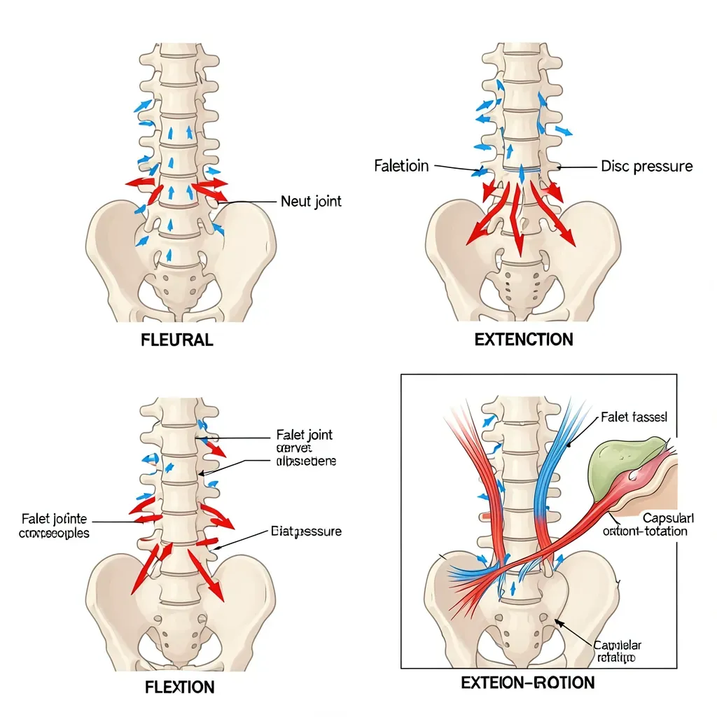
Facet joints carry approximately 20% of axial compressive load in neutral standing and substantially more in extension. When the lumbar spine moves into 15 degrees of extension, facet load increases to approximately 30% of total compressive load. At 30 degrees of extension, facet load increases further while the facet capsule simultaneously comes under tensile stress. This is why hyperextension-based exercises (aggressive back extensions, poorly coached deadlifts that extend into lumbar hyperextension) are specifically problematic for facet-dominant pain presentations.
Clinical Presentation
Facet-mediated pain has a recognizable pattern. The hallmark features, in order of diagnostic usefulness, are:
Extension and rotation aggravate symptoms. Walking uphill, standing for prolonged periods, backward bending, and lying prone all load the facets and typically worsen pain. This is the most consistent clinical finding.
Flexion relieves symptoms. Sitting (which unloads the facets by opening the posterior joint space), forward bending, and lying in the fetal position typically reduce pain. Patients often describe finding relief by "rounding forward" or by sitting down.
Morning stiffness is prominent. Facet joints accumulate fluid overnight, and the capsule is stiffened by inactivity. Pain and stiffness in the first 20-30 minutes after rising is a common facet pain feature — distinct from inflammatory arthropathy (which produces stiffness lasting hours) but longer than typical discogenic morning symptoms.
Referral is present but poorly localized. Facet joints refer pain into the buttock, posterior thigh, and occasionally the lateral calf, but this referral is diffuse and does not follow a dermatomal pattern. It does not typically radiate below the knee in a specific distribution (which would suggest nerve root involvement).
No neurological deficit. True facet pain without secondary nerve root compromise does not produce numbness, weakness, or loss of reflexes. When these features are present, additional pathology must be considered.
The extension-intolerance pattern of facet pain is easily missed when clinicians focus primarily on disc pathology. A patient whose pain consistently worsens with back extension but improves with sitting is unlikely to be primarily disc-mediated — discs are typically more painful with flexion (sitting) and more comfortable with standing and extension. Getting this distinction right is the difference between prescribing the wrong exercises and the right ones. If your pain is worse standing than sitting, worse walking uphill than flat, and is relieved by forward bending — strongly consider facet-dominant pain until proven otherwise.
Differentiating from Disc Pain
The flexion-extension pattern is the most useful clinical differentiator. As a rough rule:
- Disc-dominant pain: Worse with sitting, forward bending, and sustained flexion; better with standing, walking, and extension.
- Facet-dominant pain: Worse with standing, extension, and rotation; better with sitting and forward bending.
These patterns are not absolute. Both structures can contribute simultaneously, particularly when disc degeneration has led to facet overload. However, identifying the dominant pain generator allows exercise prescription to be tailored — which is more important than diagnostic precision for its own sake.
Diagnostic Tools: Blocks and Imaging
Medial branch blocks involve injecting a small volume of local anesthetic around the medial branch nerve supplying the suspected facet joint. If pain is significantly reduced (by convention, typically 75-80% or more) within the expected duration of the anesthetic, a facet pain diagnosis is supported. Because each facet joint is supplied by medial branches from two levels, blocks at each level are necessary. A positive block typically requires at least two confirmed responses before radiofrequency ablation is considered.
Facet joint imaging findings — joint space narrowing, subchondral sclerosis, osteophyte formation, periarticular fluid — are visible on CT and MRI but are poorly correlated with symptoms. Imaging findings guide the anatomy of injection placement but do not confirm or refute the facet as the clinical pain source in isolation.
Radiofrequency Ablation
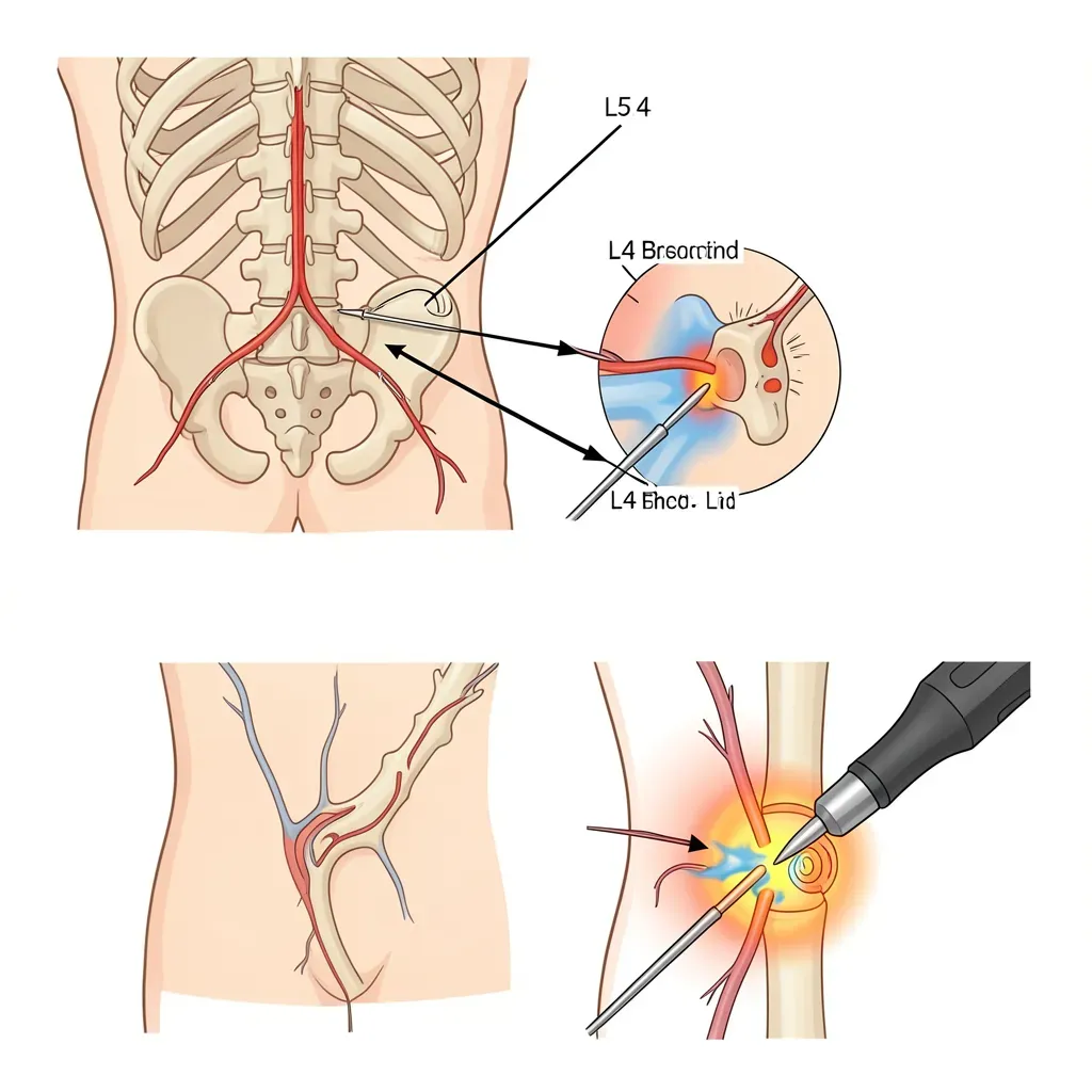
When medial branch blocks confirm the facet as the pain generator and conservative management has been inadequate, radiofrequency ablation (RFA) is a reasonable intervention. RFA uses heat generated by a radiofrequency current delivered through a needle placed adjacent to the medial branch to coagulate the nerve, interrupting pain transmission from the joint.
Outcomes for properly selected patients are good: multiple randomized controlled trials show greater pain reduction and functional improvement compared to sham procedures at 6-12 months. Duration of effect is limited — the medial branch regenerates, typically over 9-14 months, and pain may return. Repeat RFA can be performed and tends to be equally effective.
RFA is not a cure. It eliminates the pain signal from the joint without addressing the underlying joint pathology or the biomechanical factors that drove the joint to become a pain generator. Patients who use the pain relief to resume harmful movement patterns or continue the postural habits that loaded the joint excessively will return to symptomatic levels when the nerve regenerates.
Long-Term Management Through Stability Training
The most durable management for chronic facet joint syndrome involves reducing the mechanical forces on the joint during function. The key targets:
Reducing excessive lumbar extension. For people with habitual anterior pelvic tilt and exaggerated lordosis, training neutral spine position and reducing the extension range used during daily activities and exercise directly reduces facet compressive load.
Strengthening the gluteal complex. Weak gluteals are commonly associated with compensatory lumbar hyperextension during activities like climbing stairs, hip extension exercises, and single-leg stance. Strengthening hip extension through the glutes reduces the tendency to extend the lumbar spine as a compensatory strategy.
Lateral stability training. Side bridge variants, the Pallof press, and single-leg stability work reduce the rotational demands on facet joints by improving the body's ability to resist unwanted rotation through active muscular control.
The Big 3 exercises (curl-up, side bridge, bird-dog) in their standard forms are well-tolerated by facet-dominant pain presentations. The curl-up and bird-dog involve minimal lumbar extension when coached correctly. The side bridge specifically targets the lateral stability deficit common in facet pain. However, the bird-dog requires careful coaching — the arm and leg lift must not be accompanied by lumbar hyperextension or rotation, which can load an already sensitized facet joint. Start with a smaller range of motion and use a mirror or partner feedback to verify the lumbar spine remains neutral.
In Review
- Facet joints are synovial joints with rich capsular innervation; they account for 15-45% of chronic low back pain cases.
- The three failure mechanisms are acute capsular strain, osteoarthritis from cumulative loading, and chronic overload in extension postures.
- The clinical fingerprint of facet pain is extension-rotation aggravation and flexion relief — the opposite pattern to disc-dominant pain.
- Morning stiffness of 20-30 minutes and diffuse referral to the buttock and posterior thigh are characteristic; neurological deficits are absent unless nerve root compression co-exists.
- Medial branch blocks are the definitive diagnostic tool; two positive blocks at the appropriate level are required before radiofrequency ablation is considered.
- RFA provides 9-14 months of significant pain relief in properly selected patients; the medial branch regenerates and symptoms may return.
- Long-term management requires correcting the extension bias — reducing lordosis at rest, strengthening gluteals, and building lateral trunk stability.