Degenerative Disc Disease
What DDD actually means, how it differs from normal aging, why it's often a misleading diagnosis, and how to manage it.
The Problem With the Name
"Degenerative disc disease" is one of the most consequential misnomers in medicine. Strip away the alarming language and you are left with a description of something almost universal: age-related changes in the intervertebral disc that are visible on imaging.
The word "disease" implies a pathological process that is abnormal, progressive in a dangerous direction, and amenable to cure. None of those implications reliably apply to the finding of disc degeneration on an MRI. A better term — used by researchers who study this carefully — is disc degeneration, full stop. It is a description of a structural state, not a diagnosis of a disease entity.
This distinction matters enormously for recovery. People told they have a "disease" of their spine adopt catastrophic beliefs, restrict movement, and delay rehabilitation. People told their spine shows normal age-related changes — which is almost always a more accurate framing — are better positioned to do the work required.
The Thompson Grading Scale
Researchers classify disc degeneration using the Thompson grading scale, scored from I to V based on disc appearance:
Grade I: Normal. Distinct nucleus (gel-like, white on T2 MRI), intact annulus, normal disc height.
Grade II: Early degeneration. Slight darkening of nucleus, minor height reduction, annulus still intact.
Grade III: Moderate degeneration. Nucleus and annulus less clearly distinguished, moderate height loss, beginning of end plate changes.
Grade IV: Advanced degeneration. Loss of distinction between nucleus and annulus, significant height reduction, sclerotic end plates.
Grade V: Complete degeneration. Disc space collapsed, end plate erosion, nuclear material absent or solidified.
The important clinical reality: grades II and III are extremely common in adults under 50. Grade IV and V are common over 60. Most people with grades II-IV have no significant pain.
How Common Is "Abnormal" Disc Degeneration?
Population imaging studies paint a clear picture. Among people with no back pain and no symptoms:
- 30% of 30-year-olds show disc degeneration on MRI
- 50% of 40-year-olds
- 80% of 50-year-olds
- Nearly all people over 70 show some degree of disc degeneration
The finding is so prevalent that it has essentially no diagnostic value when found in isolation. A 50-year-old with back pain and disc degeneration on MRI is not distinguishable from a 50-year-old without back pain and the same imaging finding. The disc degeneration is not the explanation — it is background noise.
The pivotal question is never "does this person have disc degeneration?" Almost everyone does. The question is "does this person's pain behavior match a discogenic source, and is the imaging finding clinically correlated?" Direction of pain, aggravating positions, response to directional testing, and neurological examination matter far more than the MRI grade.
The Degeneration Cascade
When disc degeneration is clinically relevant — when it is genuinely contributing to pain rather than being an incidental finding — the mechanism follows a recognizable sequence.
Height loss: As the nucleus loses hydration, disc height decreases. This shifts load onto the annular fibers and transfers stress to the facet joints, which were not designed to be primary load-bearers.
Annular fissuring: Repeated loading of a dehydrated disc causes circumferential and radial tears in the annular fibers. The outer annulus is innervated, so fissuring can produce pain — termed internal disc disruption (IDD).
End plate changes: The vertebral end plates develop sclerosis (hardening) and Modic changes (marrow signal changes on MRI). Modic type 1 changes — inflammatory — are most consistently associated with pain. Modic type 2 — fatty replacement — are less so.
Osteophyte formation: Bone spurs form at the vertebral margins in response to abnormal motion at the degenerated segment. This is not pure damage — it is the spine's stabilization attempt.
The Stabilization Paradox
Here is the counterintuitive reality that changes how most people should think about disc degeneration: as degeneration progresses toward the advanced grades, the painful instability of the intermediate stages often resolves.
A Grade III-IV disc is mechanically unstable — enough height loss to transfer load abnormally, but not enough ankylosis (fusion) to stabilize the segment. This is typically the most symptomatic phase.
At Grade V, adjacent vertebrae may be approaching spontaneous fusion via bridging osteophytes. The segment becomes stiffer, less painful. This is why many patients with significant degenerative findings on imaging have mild or absent symptoms — their spine has already completed its stabilization process.
This means that some degree of degeneration is a destination, not a catastrophe to be reversed.
No intervention currently available reverses disc degeneration. Stem cell therapies, platelet-rich plasma injections, and disc hydration claims are not supported by high-quality evidence as of current research. Any treatment sold on the basis of "regenerating" or "reversing" your disc degeneration should be evaluated with significant skepticism. The goal of treatment is symptom management and functional restoration — not degeneration reversal.
Load Management as the Cornerstone
Because degeneration cannot be reversed, the entire clinical goal shifts to managing the load environment of the affected segment — reducing pain drivers while maintaining function.
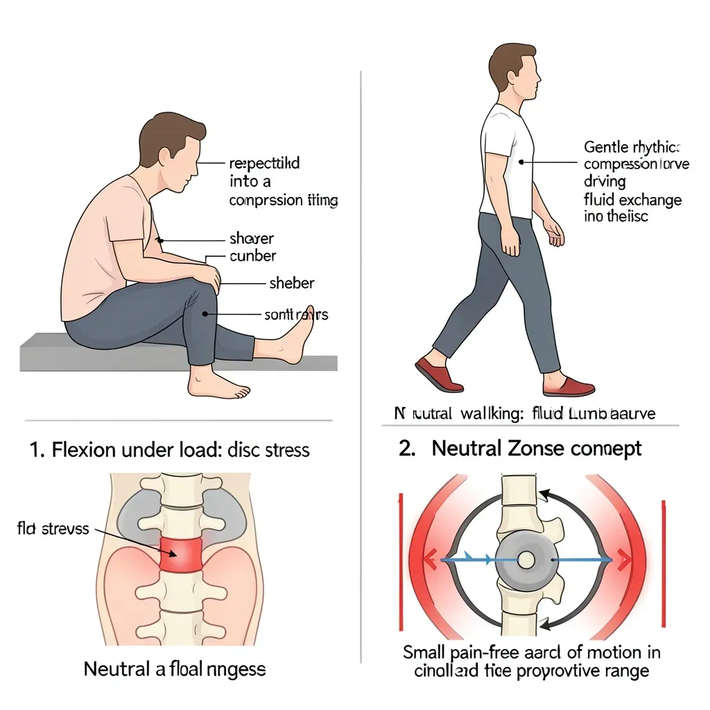
Identify your aggravating loads. Most people with discogenic pain have a clear directional bias: flexion-loaded positions (sitting, bending forward, lifting from the floor) are worse; extension is often neutral or relieving. Recognizing and temporarily reducing the aggravating direction is the first practical intervention.
Protect the neutral zone. McGill's research emphasizes that a degenerated disc has a narrowed "neutral zone" — the range of motion that can be sustained without pain. Exercises that train spinal endurance in neutral (the Big 3: curl-up, side plank, bird-dog) are the appropriate primary intervention, not stretches that push the spine into provocative ranges.
Walking remains the most accessible medicine. The compression-decompression cycle of gait pumps nutrients into degenerated discs and maintains the facet joint surfaces. A consistent daily walking program is not optional — it is the base layer of any DDD management plan.
Exercise Prescription for DDD
Exercises should be selected to build spinal endurance in neutral rather than to improve range of motion into painful directions.
Appropriate: McGill Big 3, hip hinge pattern with neutral lumbar spine (not deep forward bending), glute strengthening (bridges, clamshells), walking, swimming, cycling in an upright position.
Modify or avoid (initially): Repeated forward bending under load, loaded rotation in flexion, heavy deadlifts through full range, sit-ups and crunches, any exercise that reproduces the leg or back pain.
As the degenerated segment stabilizes and pain reduces, exercise selection can progressively expand. The goal is not permanent restriction — it is appropriate load management during the symptomatic phase, followed by graduated reloading.
Long-Term Prognosis
Population studies consistently show that back pain attributable to disc degeneration tends to improve with age. The mechanism is stabilization — the segment completes its degenerative process, osteophytes bridge across, and the hypermobility that drove pain resolves into stiffness.
This is not a guarantee of a pain-free future — it is context that should reduce catastrophizing. Most people with DDD who manage their loads intelligently, maintain fitness, and address the psychological dimensions of pain do well over the long term.
The single most powerful predictor of long-term outcome in DDD is not the imaging grade or the disc level — it is whether the person remains physically active. Deconditioning, weight gain, and movement avoidance accelerate the downward spiral. Staying strong, staying lean, and maintaining the walking habit is the best evidence-based treatment protocol available.
In Review
- "Degenerative disc disease" is a description of age-related disc changes, not a true disease — the name is misleading and drives unnecessary fear
- The Thompson grading scale (I-V) describes disc degeneration severity; grades II-III are nearly universal in adults over 40
- 30% of asymptomatic 30-year-olds have disc degeneration on MRI; imaging findings must be clinically correlated, not treated as automatic diagnoses
- The degeneration cascade — height loss, annular fissuring, end plate changes, osteophyte formation — can produce pain in its intermediate stages but often stabilizes with age
- No treatment reverses disc degeneration; the goal is load management and symptom control
- Exercise in neutral spine, walking, and load management are the cornerstone interventions
- Long-term prognosis is often favorable as the segment stabilizes — remaining physically active is the strongest predictor of good outcome