Thoracic Spine Pain: The Neglected Middle Back
Understanding pain between the shoulder blades — facet joint referral, costovertebral dysfunction, postural drivers, and why thoracic mobility matters for the whole spine.
The Forgotten Region
The thoracic spine — the twelve vertebrae that make up the mid-back, attached to the rib cage — receives far less clinical attention than the lumbar spine and neck. Most back pain research, most rehabilitation programs, and most clinical guidelines focus on the lower back. Yet pain between the shoulder blades is among the most common musculoskeletal complaints, particularly in people who spend long hours at a desk or in sustained seated postures.
Thoracic pain is also clinically important beyond the discomfort it causes directly. The thoracic spine is the mechanical foundation of the entire spinal column. When it becomes stiff — as it reliably does in people with sedentary lifestyles — the lumbar spine and cervical spine compensate, leading to pain and dysfunction at those levels. Understanding thoracic pain therefore means understanding how the thoracic spine affects the whole system.
Anatomy: The Thoracic Spine Is Not Just Vertebrae
The thoracic spine is structurally unique because each vertebra articulates with the rib cage. Each rib connects to its corresponding thoracic vertebra at two points: the costovertebral joint, where the rib head meets the vertebral body, and the costotransverse joint, where the rib contacts the transverse process. This dual articulation means there are far more joints in the thoracic region than in the lumbar spine, and any of them can become a pain generator.
The facet joints of the thoracic spine are oriented at approximately 60 degrees from horizontal — compared to roughly 90 degrees (near-vertical) in the lumbar spine. This orientation makes the thoracic facets primarily designed to resist rotation, which makes sustained rotation or end-range rotation the primary mechanical stressor for thoracic facets.
The thoracic spinal cord is fully contained within the thoracic canal, and the thoracic nerve roots exit quite specifically. Thoracic radiculopathy — nerve root irritation in the thoracic region — can produce band-like pain wrapping around the chest wall, which is a recognized but underdiagnosed cause of mid-back and chest pain.
Costovertebral Joint Dysfunction
Costovertebral joints are small, synovial joints that move with every breath. Inspiration expands the rib cage; expiration compresses it. Over the course of a day, these joints move thousands of times. When they become restricted or irritated — often from sustained forward-bent posture, repetitive thoracic flexion, or direct trauma — the pain can be remarkably localized and reproducible.
The clinical presentation of costovertebral dysfunction typically includes pain precisely localized to one or two levels of the mid-back, often just lateral to the spine. It is often provoked by deep breathing, twisting, reaching overhead, or pressure applied directly to the rib angle. It frequently has a sharp, catching quality and may refer anteriorly around the chest wall in a dermatomal or non-dermatomal pattern.
Because costovertebral pain can refer to the chest wall, it is important to recognize when chest pain has a musculoskeletal origin versus a cardiac or pulmonary one. If thoracic or chest pain is reproduced by palpation of the rib-spine junction, changes with breathing but not with exertion, and has been present for weeks or months in a young or middle-aged person with no cardiac risk factors, a musculoskeletal origin is likely. When in doubt, cardiac causes should always be excluded first.
Thoracic and mid-back pain that is accompanied by shortness of breath at rest, chest tightness with exertion, sweating, arm or jaw pain, or pain that is progressive and unrelenting should be evaluated medically without delay. These are not features of mechanical thoracic spine pain and require exclusion of cardiac or pulmonary pathology.
Thoracic Facet Referral Patterns
The thoracic facet joints refer pain in patterns that are less predictable than lumbar facet referral but nonetheless recognizable. Upper thoracic facet joints (T1-T4) tend to refer into the posterior shoulder, neck, and upper arm. Mid-thoracic joints (T4-T8) refer locally to the mid-back and can refer anteriorly to the chest wall. Lower thoracic joints (T8-T12) refer to the lower thoracic region and can mimic lumbar pain or even abdominal pain.
The challenge with thoracic facet referral is that it can closely mimic other conditions: upper thoracic referral mimics cervical radiculopathy or rotator cuff pathology; lower thoracic referral mimics lumbar pain or internal organ pathology. A careful history — including what makes the pain better and worse — combined with palpation and mobility testing is often the most reliable way to identify the thoracic facets as the pain source.
Thoracic Kyphosis and Its Compensations
The thoracic spine normally has a kyphotic curve — a gentle forward bend of approximately 20–45 degrees. This is a structural feature, not a pathology. However, when thoracic kyphosis becomes excessive — beyond roughly 50–55 degrees — the mechanical consequences cascade through the entire spine.
An excessively kyphotic thoracic spine forces the cervical spine into extension (the head must remain level, so the neck extends over the rounded upper back). This compresses the cervical facets and can contribute to cervical facet pain, cervicogenic headaches, and upper trapezius pain. Simultaneously, the excessively kyphotic thoracic spine limits thoracic extension during movements that require it — reaching overhead, throwing, looking up. The lumbar spine then extends beyond its optimal range to compensate, loading the lumbar facets and posterior elements.
This compensation cascade is why thoracic stiffness is a lumbar pain driver. The lumbar spine is not primarily responsible for thoracic problems, but it pays the mechanical price for them. Patients presenting with lumbar facet pain and no resolution despite appropriate lumbar treatment should always be assessed for thoracic mobility deficits.
A simple clinical screen for thoracic mobility contribution to lumbar pain: have the patient sit on a chair (eliminating hip mobility from the equation) and rotate the thoracic spine to each side. Restriction or pain with this movement, combined with a clinical picture of lumbar pain, strongly suggests the thoracic spine is contributing. Normal thoracic rotation from a seated position is approximately 35–50 degrees per side.
Rib Cage Mechanics and Breathing
The rib cage is a dynamic structure that changes volume with every breath. Full thoracic expansion requires that the ribs move upward and outward (bucket handle motion) and forward (pump handle motion) with inspiration. When the thoracic spine is stiff, these rib movements are restricted, breathing becomes shallower, and the accessory breathing muscles — scalenes, sternocleidomastoid, pectorals — take on a larger share of the respiratory work. This creates chronic tension in the neck, shoulder girdle, and upper thoracic region.
Conversely, improving thoracic mobility often has an immediate and noticeable effect on breathing depth and ease. Patients who perform a thoracic extension mobilization session over a foam roller frequently report not only reduced mid-back pain but also a sense of being able to breathe more freely. This is not coincidental — restoring rib cage mobility directly improves respiratory mechanics.
Sustained Desk Posture: The Primary Driver
The most common cause of thoracic stiffness and pain in otherwise healthy people is sustained seated posture with thoracic flexion. Sitting at a desk with a forward head, rounded upper back, and protracted shoulder girdle for six to eight hours a day reliably produces thoracic extension stiffness, costovertebral joint restriction, and the compensatory patterns described above.
The mechanics are straightforward: sustained flexion compresses the anterior vertebral bodies and disc spaces, places the posterior facet joints in a distracted position, and allows the posterior thoracic musculature to remain in a lengthened, eccentrically loaded position for hours. Over time, this produces adaptive shortening of the anterior thoracic structures (pectorals, intercostals, anterior longitudinal ligament), loss of thoracic extension range, and the classic upper crossed syndrome pattern of forward head posture.
The treatment principle follows directly: if the problem is sustained flexion, the solution is extension-biased mobility work, postural loading variation, and correction of the sustained positions that are driving the problem.
Thoracic Extension Exercises
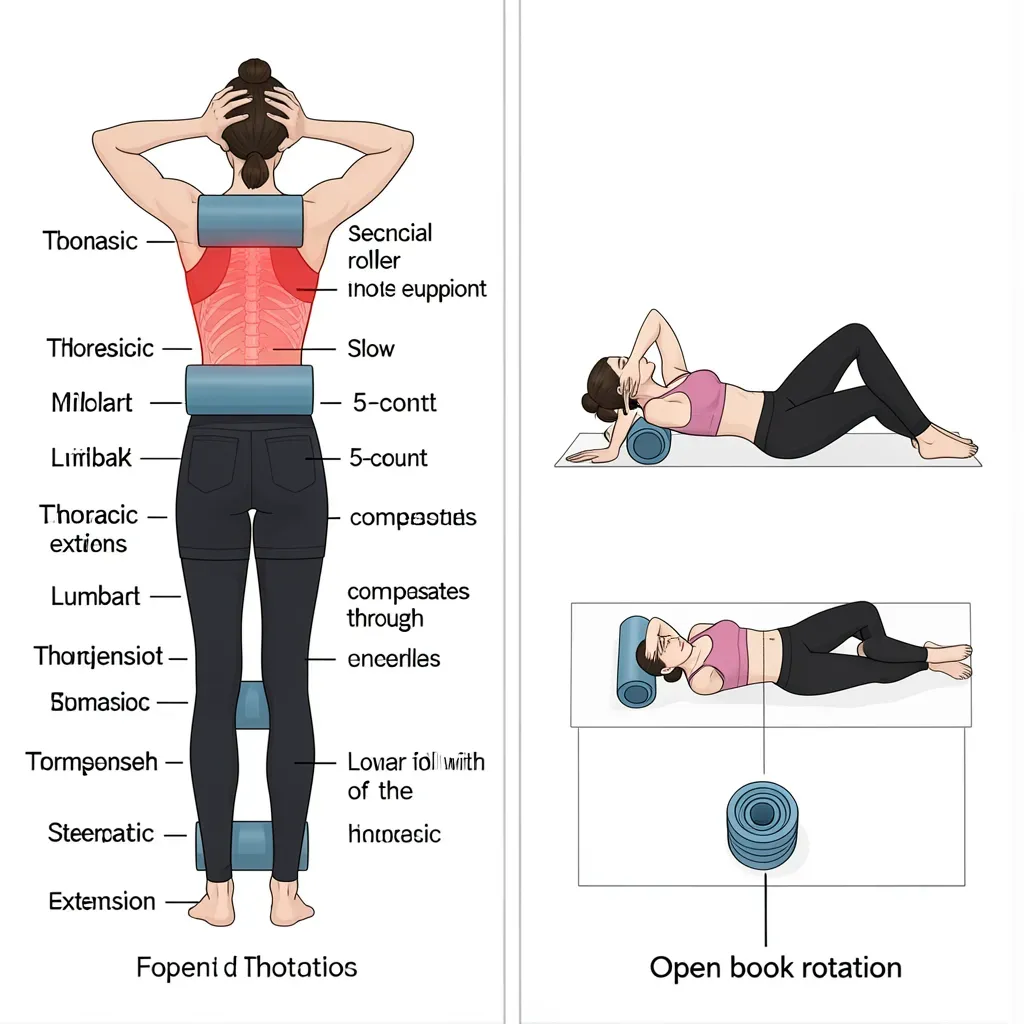
The thoracic foam roller extension is the most practical and widely accessible thoracic extension exercise. The foam roller is placed perpendicular to the spine at one thoracic level; the patient lies back over it with hands behind the head for support. The weight of the head and upper body gently extends the thoracic spine over the apex of the roller. The key is segmental specificity: the roller should be moved to each thoracic level, spending 5–8 slow extension repetitions per level from approximately T4 to T10. Avoid placing the roller at the lumbar spine, which does not benefit from extension mobilization in the same way.
A second effective exercise is the open book rotation: lying on the side with hips and knees at 90 degrees, the top arm sweeps upward and back toward the floor behind, rotating the thoracic spine into extension and rotation. This targets the same muscles and joints that are stiffened by prolonged forward posture.
For strengthening the thoracic extensors, the prone Y and T exercises are highly effective. Lying prone with arms raised in a Y or T position, the patient lifts the arms off the floor by engaging the lower trapezius and mid-thoracic extensors. These muscles are typically weak in people with thoracic kyphosis and are direct contributors to posture and rib cage mechanics.
When Thoracic Pain Is Serious
Most thoracic pain is mechanical and responds to the interventions described above. However, the thoracic spine is also a location where serious pathology — including metastatic cancer, fractures, and aortic pathology — can first present as back pain. Several features should prompt medical evaluation rather than self-treatment:
- Pain that is constant, progressive, and unrelated to movement or position
- Night pain that wakes the patient from sleep and does not resolve when they move around
- Pain in a person over 50 with a history of cancer, even a remote one
- Thoracic pain accompanied by lower extremity weakness, numbness, or difficulty walking (possible thoracic cord compression)
- Acute onset of severe thoracic pain in a postmenopausal woman or older man (possible compression fracture)
- Pain accompanied by fever, unexplained weight loss, or systemic symptoms
These are red flags. Mechanical thoracic pain fluctuates with movement, is better in some positions than others, and has a recognizable provocation pattern. Pain that does not behave this way requires investigation.
In Review
- The thoracic spine articulates with the rib cage at costovertebral and costotransverse joints, providing far more potential pain generators per spinal level than the lumbar spine
- Costovertebral joint dysfunction presents as sharp, localized mid-back pain provoked by breathing and rotation, and can refer anteriorly around the chest wall
- Thoracic facet joints refer pain into the posterior shoulder and neck (upper thoracic), chest wall (mid-thoracic), and lower back or abdomen (lower thoracic)
- Excessive thoracic kyphosis forces cervical extension compensation and lumbar hyperextension, making thoracic stiffness a primary driver of both neck and lumbar pain
- Sustained seated posture with thoracic flexion is the dominant cause of thoracic stiffness in desk workers; the treatment principle is extension-biased mobility and postural loading variation
- Thoracic foam roller extension and open book rotation are the most practical and evidence-aligned exercises for restoring thoracic extension mobility
- Breathing mechanics depend on rib cage mobility; improving thoracic extension often produces immediate improvements in respiratory depth and ease
- Red flags for serious thoracic pathology include constant progressive pain, night pain, history of cancer, and neurological symptoms — these require medical evaluation rather than exercise