Back Pain and Diabetes: Shared Mechanisms and Complications
How diabetes accelerates disc degeneration and peripheral neuropathy, and how the same lifestyle interventions improve both conditions.
Why Diabetes and Back Pain Converge
Type 2 diabetes and chronic back pain share more biological terrain than most patients — or their physicians — realize. They co-occur at higher rates than chance predicts, they share overlapping pathophysiological mechanisms, and critically, the most evidence-based interventions for each condition overlap substantially. This convergence is not coincidental: both conditions are downstream consequences of the same upstream drivers — insulin resistance, chronic systemic inflammation, and metabolic dysfunction.
The clinical significance is practical. A patient managing both conditions does not need two entirely separate treatment programs. The exercise protocols, dietary changes, and lifestyle modifications that improve glycemic control also improve pain outcomes. The same inflammation-reducing dietary changes that benefit spinal tissue health improve insulin sensitivity. Understanding the specific mechanisms connecting diabetes to disc health helps patients appreciate that managing their metabolic health is directly managing their back pain — not just a separate health priority.
Advanced Glycation End Products and Disc Tissue
The most direct and specific mechanism connecting diabetes to accelerated disc degeneration is the accumulation of advanced glycation end products (AGEs) in disc tissue.
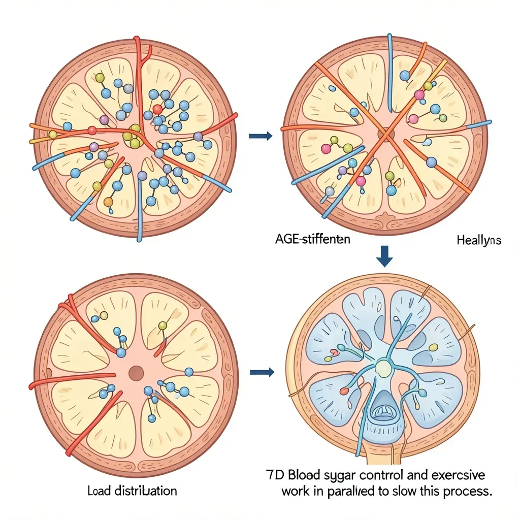
AGEs form when glucose molecules spontaneously bind to proteins and lipids through non-enzymatic glycation reactions — the same chemistry that browns toast. This process occurs in all humans, but it accelerates dramatically in high-glucose environments. In people with chronic hyperglycemia, AGE accumulation occurs at an accelerated rate in long-lived structural proteins — collagen, elastin, and the proteoglycans of the intervertebral disc are primary targets.
When AGEs cross-link collagen and disc matrix proteins, they produce a structural stiffening of the tissue. The disc nucleus pulposus normally has high water content and viscoelastic properties that allow it to distribute compressive loads by bulging radially. AGE cross-linking of disc matrix proteins reduces this hydration capacity and viscoelastic deformability — the disc becomes stiffer, loses height faster, and transmits compressive loads with less energy absorption. The result is mechanical stress concentration at the annulus fibrosus and the vertebral endplates, predisposing to annular tears, endplate lesions, and accelerated degenerative cascade.
This is not an indirect or speculative mechanism. Post-mortem studies have directly measured AGE concentration in disc tissue and found strong correlations with disc degeneration severity. In diabetic populations, disc AGE content is elevated above age-matched controls, paralleling the accelerated disc degeneration seen on imaging in diabetic patients.
Peripheral Neuropathy vs. Nerve Root Compression: A Critical Differential
One of the most clinically significant diagnostic challenges at the intersection of diabetes and back pain is distinguishing diabetic peripheral neuropathy (DPN) from lumbar radiculopathy. Both conditions produce leg pain, tingling, numbness, and weakness. The consequences of misdiagnosis are significant — a patient with DPN who receives spinal surgery for presumed radiculopathy will not improve from the surgery, while a patient with actual radiculopathy may miss appropriate treatment if their symptoms are attributed entirely to neuropathy.
Diabetic peripheral neuropathy characteristics:
- Stocking-and-glove distribution — begins in the toes and feet, advances proximally and symmetrically
- Burning, tingling, or "electric" quality
- Worse at rest and at night (a distinguishing feature — radicular pain is typically worse with loading and better with rest)
- Associated with vibration sense loss, reduced ankle deep tendon reflexes
- Electrodiagnostic studies (EMG/nerve conduction velocity) show peripheral nerve involvement with characteristic DPN pattern
Lumbar radiculopathy characteristics:
- Dermatomal distribution — corresponds to specific nerve root territory (L4: medial shin, L5: lateral shin and dorsal foot, S1: lateral foot and heel)
- Often sharp, shooting quality down the leg
- Worse with spinal loading, sitting, or positions that increase disc pressure (Valsalva maneuver)
- Better with lying down and unloading
- Specific positive provocative tests (straight leg raise, slump test)
The two conditions can coexist, which is the most diagnostically challenging scenario. A diabetic patient with pre-existing DPN who develops a new disc herniation may have both simultaneously — the neuropathy changes the baseline sensation, making the additional radicular component harder to identify. In ambiguous cases, MRI with nerve root correlation, electrodiagnostic testing, and selective nerve root blocks are all useful for delineating the respective contributions.
Insulin Resistance and Systemic Inflammation: The Shared Pathway
Insulin resistance — the reduced cellular responsiveness to insulin that characterizes type 2 diabetes and its precursor state — is not merely a glucose regulation problem. It is fundamentally a pro-inflammatory state that produces elevated circulating levels of the same cytokines that drive disc and joint inflammation: TNF-α, IL-6, IL-1β, and C-reactive protein.
The mechanism runs through visceral adipose tissue (covered in more detail in chapter 117), which in insulin-resistant states becomes a major source of inflammatory adipokines. Elevated TNF-α specifically contributes to the spine pain pathway by directly sensitizing the nociceptors in disc tissue — disc material that has leaked through annular tears becomes highly inflammatory when it contacts neural tissue, and this inflammatory effect is amplified in a high-TNF-α systemic environment.
The shared inflammatory pathway means that interventions that reduce insulin resistance also reduce the systemic inflammatory environment that amplifies back pain. This creates a treatment multiplier: improving glycemic control is simultaneously managing spine inflammation. The same 5–10% weight reduction that meaningfully reduces inflammatory cytokines in type 2 diabetes patients also reduces spinal inflammatory burden and disc nociceptor sensitization.
Exercise: Doubly Beneficial
The evidence for exercise as a primary intervention in type 2 diabetes is as strong as the evidence for any pharmaceutical. Structured exercise — both aerobic and resistance training — improves insulin sensitivity directly and independently of weight loss through multiple mechanisms: increased glucose transporter (GLUT-4) translocation to muscle cell membranes, reduced ectopic lipid deposition in muscle tissue, improved mitochondrial density and fatty acid oxidation capacity, and reduced hepatic glucose output.
For back pain specifically, exercise is the single most evidence-supported intervention across all guidelines. The mechanisms are discussed throughout this guide: improved disc nutrition through cyclic loading, improved paraspinal and core muscle support, reduced fear-avoidance behavior, and improved sleep quality that supports disc rehydration.
The combination creates an unusual situation where the same exercise prescription addresses both conditions simultaneously. A patient who performs three to four sessions per week of moderate-intensity aerobic exercise (walking, cycling, swimming) combined with two sessions of resistance training is following the optimal program for both glycemic control and back pain management. The resistance training component — often underemphasized in diabetes management programs — is particularly relevant because muscle mass is the primary site of insulin-stimulated glucose disposal.
The specific adaptations are additive: exercise improves insulin sensitivity, reducing glycemic peaks and HbA1c. Reduced hyperglycemia slows AGE accumulation in disc tissue. Reduced disc AGE accumulation slows the structural stiffening and hydration loss that accelerate degeneration. Meanwhile, the mechanical benefits of exercise (muscle support, disc nutrition) work in parallel. A patient who exercises consistently is addressing the disc through both the mechanical and metabolic pathways.
Blood Sugar Control and Pain Threshold
Acute hyperglycemia has effects on pain threshold beyond the chronic AGE accumulation mechanism. Elevated blood glucose promotes oxidative stress (excess reactive oxygen species from glucose auto-oxidation), which directly sensitizes nociceptors in peripheral tissue. Studies have shown that people in hyperglycemic states have lower pain thresholds and higher pain ratings from the same experimental stimulus compared to their euglycemic baseline.
This means that glycemic excursions — the blood sugar spikes after high-glycemic meals — produce immediate, measurable increases in pain sensitivity. For a back pain patient who eats a high-glycemic meal and then notices that their back pain seems worse in the subsequent hours, this is not coincidence or psychosomatic response — it reflects a real, physiologically mediated change in peripheral nociceptor sensitivity.
The practical implication: glycemic variability management, not just average HbA1c, is clinically relevant to pain outcomes. Reducing post-meal glucose spikes through lower-glycemic food choices, meal composition (protein and fat slow glucose absorption), and physical activity after eating (a 10–15 minute walk post-meal meaningfully reduces the post-prandial glucose spike) can reduce the frequency and severity of pain excursions.
Medication Effects on Pain
Metformin — the first-line pharmaceutical for type 2 diabetes — has a neutral to potentially positive profile for pain. Some research has found metformin has direct anti-inflammatory properties through AMPK activation and NF-κB inhibition. Its primary clinical value for back pain patients is its metabolic effect: by improving insulin sensitivity and reducing inflammatory markers, metformin's therapeutic benefit also partially reduces the pro-inflammatory state driving pain amplification.
GLP-1 receptor agonists (semaglutide, liraglutide, tirzepatide) — the most effective weight-loss medications currently available, also effective for glycemic control. Through their weight reduction effect (covered in chapter 117), they reduce spinal compressive load, visceral adiposity-derived inflammation, and mechanical strain from adipose-related anterior pelvic tilt. Some direct anti-inflammatory effects of GLP-1 agonism are also described independent of weight loss.
Thiazolidinediones (pioglitazone, rosiglitazone) — PPAR-gamma agonists with direct anti-inflammatory properties in addition to insulin-sensitizing effects. Some evidence suggests beneficial effects on inflammatory pain independent of glycemic effects.
Sulfonylureas and older insulin secretagogues — do not have evidence for direct pain benefit, and the hypoglycemic events they can produce (low blood sugar) may actually increase pain sensitivity acutely.
In Review
- AGE accumulation in disc matrix proteins is the direct mechanism by which chronic hyperglycemia accelerates disc degeneration — it stiffens the disc, reduces hydration, and increases mechanical stress concentration
- Endplate AGE cross-linking also reduces nutrient diffusion into the avascular disc, creating a secondary degeneration pathway
- Distinguishing diabetic peripheral neuropathy from lumbar radiculopathy is essential — both produce leg symptoms but require different treatments; they frequently coexist
- The stocking-and-glove distribution, night-dominant pattern, and bilateral symmetry of DPN differ from the dermatomal, load-worsened pattern of radiculopathy
- Insulin resistance drives elevated TNF-α, IL-6, and IL-1β — the same cytokines that sensitize disc nociceptors and amplify pain
- Exercise improves insulin sensitivity and disc health simultaneously — the same program addresses both conditions in parallel
- Acute hyperglycemia reduces pain threshold through oxidative stress and nociceptor sensitization — post-prandial glycemic control matters for daily pain experience
- Metformin and GLP-1 agonists have favorable profiles for back pain patients through their anti-inflammatory and weight-reduction effects respectively