Cervical Spine Pain
Neck pain, cervical disc herniation, and cervical stenosis — the same biomechanical principles applied to the upper spine.
The Cervical Spine: Same Principles, Different Details
The lumbar and cervical spines share fundamental architecture — vertebral bodies, intervertebral discs, facet joints, nerve roots exiting through foramina — and the biomechanical principles that govern lumbar pain apply with important modifications to the cervical spine.
The cervical spine has seven vertebrae (C1-C7). C1 (atlas) and C2 (axis) are atypical — the atlas is a bony ring that supports the skull, and the axis has the odontoid process (dens) around which the atlas rotates. Together they account for approximately 50% of cervical rotation. C3 through C7 are more conventional mobile segments with true intervertebral discs, facet joints, and foramina through which nerve roots exit.
The facet joints in the cervical spine are oriented more horizontally than lumbar facets, allowing the neck its substantial range of motion — flexion, extension, lateral bending, and rotation — but making it more vulnerable to shear forces. The intervertebral discs are smaller relative to the vertebral bodies than lumbar discs, and the uncinate processes (raised bony lips on the posterolateral corners of each vertebral body) partially constrain lateral disc herniation — shunting herniations toward the posterior-lateral direction.
The Most Common Herniation Levels
C5-C6: The most common cervical herniation level. The C6 nerve root is affected. Classic referral pattern: pain from the neck, across the top of the shoulder, down the lateral arm, and into the thumb and index finger. Biceps reflex may be reduced. Wrist extension weakness (extensor carpi radialis) is possible.
C6-C7: The second most common level. The C7 nerve root is affected. Referral pattern: pain from the neck into the posterior arm and into the middle finger. Triceps reflex may be reduced. Triceps weakness is possible.
C4-C5: Less common. C5 nerve root. Pain and potential weakness in the deltoid and shoulder external rotators. May be confused with shoulder pathology.
C7-T1: Uncommon. C8 nerve root. Pain into the ring and little fingers, intrinsic hand weakness — important to distinguish from ulnar nerve compression at the elbow.
The arm pain from cervical radiculopathy typically has a burning, electric quality along the specific dermatomal distribution. It is often worse with neck extension and ipsilateral lateral bending (which closes the foramen and compresses the nerve root) and improved by placing the arm overhead or behind the head (which opens the foramen — the Bakody sign).
The Spurling test (cervical extension + lateral bending toward the painful side + axial compression) reproduces radicular arm pain in cervical disc herniation. A positive Spurling has high specificity for cervical radiculopathy — if it provokes the familiar arm pain, the diagnosis is very likely correct. A negative Spurling does not rule it out, as sensitivity is lower.
Text Neck and Forward Head Posture
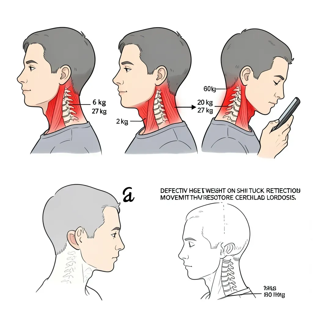
The human head weighs approximately 5-6 kilograms in neutral alignment. As the head migrates forward of the center of gravity — as it does with prolonged phone use, laptop work, and driving — the effective load on the cervical spine increases dramatically:
- At 15 degrees of forward head tilt: approximately 12 kg
- At 30 degrees: approximately 18 kg
- At 45 degrees: approximately 22 kg
- At 60 degrees: approximately 27 kg
The sub-occipital and posterior cervical muscles must continuously contract to prevent the head from falling further forward. This sustained isometric contraction produces the deep, aching cervical and upper trapezius pain that is epidemic in desk workers and phone users.
Forward head posture also reduces the cervical lordosis (the normal backward curve of the neck), which increases disc pressure in the posterior cervical segments and narrows the neuroforamina — predisposing to nerve root irritation over time.
Cervical Extension Exercises: The McKenzie Approach
The directional preference principle that underlies lumbar rehabilitation applies equally to the cervical spine. Most cervical disc herniations are aggravated by flexion (which loads the posterior disc and opens the anterior vertebral spacing, pushing the nucleus posteriorly) and relieved by extension (which reverses the nuclear migration).
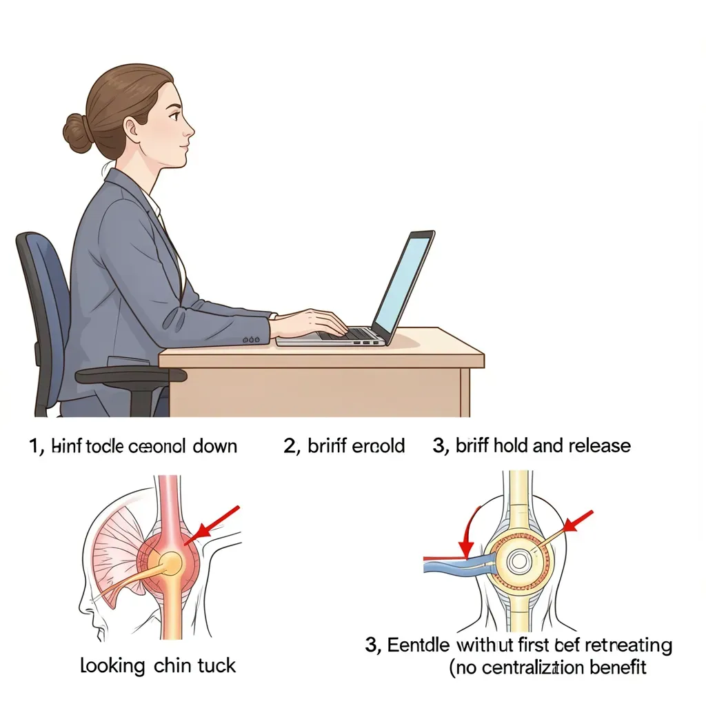
Chin tuck (cervical retraction): The cornerstone cervical exercise. Draw the chin straight backward — not downward — as if making a "double chin." This movement restores cervical lordosis, retracts the head over the shoulders, and loads the posterior disc in a way that favors nuclear centralization. Hold for 5-10 seconds, repeat 10 times, perform every 1-2 hours.
Cervical extension in retraction: From the chin-tuck position, gently extend the neck backward. This is McKenzie's cervical extension exercise. Start small and progressively increase range over days as symptoms permit. If arm pain or neurological symptoms increase, stop and reassess.
Cervical lateral flexion and rotation: Mobility exercises in these planes are appropriate once acute nerve root irritation has settled. They should not be the primary intervention in acute radiculopathy.
The centralization principle works in the cervical spine exactly as it does in the lumbar spine. If performing cervical retraction and extension causes arm pain to move toward the neck (centralize), this is a positive prognostic sign — continue the exercises. If arm pain moves further down the arm (peripheralizes), discontinue that direction and reassess. The movement direction that produces centralization is the treatment.
Cervical Stenosis and Myelopathy
Cervical stenosis — narrowing of the spinal canal in the cervical spine — is more clinically significant than lumbar stenosis because the cervical canal contains the spinal cord, not just the cauda equina.
Cervical myelopathy (spinal cord compression) is a surgical emergency when the following signs are present:
- Progressive hand weakness or clumsiness (difficulty buttoning shirts, dropping objects)
- Gait deterioration — "walking like I'm drunk," wide-based or unsteady gait
- Hoffman's sign (flicking the middle finger nail produces reflex flexion of the thumb — upper motor neuron sign)
- Hyperreflexia in the lower extremities
- Lhermitte's sign — electric shock sensation down the spine and limbs with neck flexion
These signs indicate cord compression that is causing upper motor neuron dysfunction. Unlike nerve root compression (radiculopathy), which can be managed conservatively in most cases, established myelopathy requires surgical decompression to prevent permanent cord damage. If you have neck pain accompanied by any of the above — seek urgent neurological assessment.
Cervical stenosis without myelopathy (cord contact but no cord dysfunction) can often be managed conservatively with modification of provocative movements, activity guidance, and exercise — similar to lumbar stenosis.
The Upper Cervical Spine and Manipulation
A note on cervical manipulation: evidence supports cervical manipulation for neck pain and headaches arising from the cervical spine. However, high-velocity manipulation of the upper cervical spine (C1-C2) carries a rare but documented risk of vertebral artery dissection, which can cause stroke. The estimated rate is extremely low — approximately 1 in 1-2 million procedures — but the consequence of the rare adverse event is severe.
For the vast majority of people with cervical disc herniation and radiculopathy, high-velocity manipulation is contraindicated regardless of vertebral artery risk, as it is mechanically unsuitable for a nerve root already under compressive stress. Mobilization (sustained, low-velocity), cervical retraction exercises, and traction are safer and evidence-supported alternatives.
Exercises That Rebuild Cervical Resilience
Long-term cervical spine health requires more than extension exercises. The deep cervical flexors — longus colli and longus capitis — are the cervical equivalent of the lumbar multifidus: small, deep stabilizers that are the first to inhibit and atrophy in cervical pain conditions.
Deep cervical flexor training (craniocervical flexion test pattern): Lying supine, perform a gentle nodding action — yes-nodding at the craniocervical junction, not a full neck curl-up. Pressure biofeedback targets 22-26 mmHg above baseline. This exercise is highly specific to the deep stabilizers and is the cervical equivalent of the transverse abdominis activation that underlies lumbar stability.
Scapular setting: Retracting and depressing the scapulae reduces upper trapezius over-recruitment, reduces forward head posture, and takes compressive stress off the lower cervical spine.
Thoracic extension: Restoring thoracic extension mobility directly reduces cervical compensatory posturing. If the mid-back is kyphotic and stiff, the cervical spine hyperextends or forward-protrudes to maintain the visual horizon. Thoracic extension exercises (foam roller thoracic extension, book openings) have a direct positive effect on cervical mechanics.
In Review
- C5-C6 and C6-C7 are the most common cervical herniation levels, producing C6 (thumb/index finger) and C7 (middle finger) radiculopathy respectively
- Forward head posture multiplies cervical load dramatically — 6 kg neutral becomes 27 kg at 60 degrees of forward tilt
- The McKenzie directional preference principle applies to the cervical spine — chin tuck and cervical extension produce centralization for most disc herniations
- Cervical myelopathy (hand clumsiness, gait changes, Hoffman's sign, hyperreflexia) is a surgical emergency — seek urgent assessment
- High-velocity upper cervical manipulation is contraindicated for disc herniation radiculopathy; mobilization and retraction exercises are appropriate
- Deep cervical flexor training and thoracic extension are essential components of cervical rehabilitation that extend beyond symptom management