SI Joint Dysfunction
How to identify sacroiliac joint pain, distinguish it from lumbar pain, and use targeted stabilization strategies.
Anatomy of the Sacroiliac Joint
The sacroiliac joint (SIJ) connects the sacrum — the triangular bone at the base of the spine — to the iliac wings of the pelvis on each side. It is one of the largest joints in the body and, under normal circumstances, one of the least mobile.
The SIJ transmits all the forces between the upper body and the lower extremities. In upright stance, this means the entire weight of the torso, plus any load carried, passes through both SIJs with every step. The joint is designed for stability, not mobility. Its irregular, interlocking surfaces and extremely dense surrounding ligament system (the posterior sacroiliac ligaments are among the strongest in the body) reflect this priority.
Normal SIJ motion is small — measured in millimeters and fractions of a degree. The joint does move during certain activities (notably the last stages of pregnancy and delivery), but it is not designed as a range-of-motion contributor to normal movement. When it is perceived as "stuck" or "out of place," the clinical problem is almost always not reduced mobility but reduced stability.
The concept of the SIJ being "out of alignment" and requiring manipulation to "put it back" is mechanically implausible given the joint's anatomy. The interlocking surfaces and surrounding ligament density make significant malalignment virtually impossible without major trauma. What is being treated when SIJ manipulation provides relief is likely an articular reflex or temporary pain modulation — not a structural realignment.
Why SI Joint Pain Is Commonly Misdiagnosed
The SIJ produces pain that mimics — often precisely — lumbar disc herniation and L5-S1 radiculopathy. The reasons:
- Pain is typically located in the posterior pelvis, directly over or just below the posterior superior iliac spine (PSIS)
- Pain commonly refers to the buttock, groin, and posterior thigh
- It can refer down the leg, though rarely below the knee (unlike true radiculopathy, which consistently produces below-knee symptoms)
- L5-S1 disc herniation affects the same region
The result: SIJ dysfunction is frequently treated as lumbar disc disease, sometimes with surgery, often with failure. Accurate diagnosis requires specific provocation testing, not imaging alone.
Imaging (X-ray, CT, MRI) is useful for ruling out sacroiliac arthropathy, infection, or inflammatory disease (ankylosing spondylitis). But in mechanical SIJ dysfunction, imaging is typically normal or shows only non-specific degenerative change. The diagnosis is clinical.
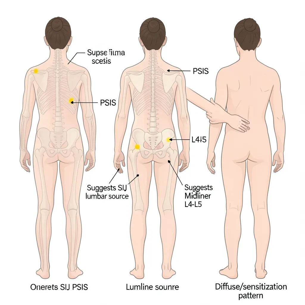
The One-Finger Test
A remarkably useful, simple screening tool: ask the patient to point with one finger to the location of their worst pain.
- SIJ pain: The patient points to a small, focal area directly over or just medial to the PSIS — a specific bony landmark you can identify by palpating the posterior iliac crest and following it to its posterior projection
- Lumbar pain: The patient points to the midline or paramidline of the low back, typically above the level of the PSIS
- Referred/vague pain: The patient gestures broadly across the low back and buttock with an open hand — suggesting either sensitization, non-specific pain, or difficulty localizing
The one-finger test doesn't confirm SIJ dysfunction — it suggests where to direct clinical testing.
Provocation Tests
No single test confirms SIJ dysfunction. Clinical confidence comes from finding multiple positive provocation tests that collectively point to the SIJ.
FABER test (Patrick's test): Patient lies supine. Examiner places one leg in Flexion, ABduction, and External Rotation — the figure-four position. Gentle downward pressure on the knee. Positive if pain is reproduced in the ipsilateral posterior pelvis (over the SIJ). Note: hip pathology can also produce a positive FABER — location of reproduced pain distinguishes them.
Gaenslen's test: Patient lies supine at the edge of the table. One leg hangs off the edge (hip extended), the other is pulled to the chest (hip flexed). The counter-rotation between the two hemi-pelves stresses the SIJ on the hanging-leg side. Positive if ipsilateral posterior pelvic pain is reproduced.
Thigh thrust test: Patient supine. Examiner flexes the hip to 90 degrees and applies an axial force down through the femur toward the SIJ (posterior shear force on the sacrum relative to the ilium). Positive if posterior pelvic pain is reproduced. This is the most specific single test for SIJ pain.
Compression test: Patient side-lying. Examiner applies a compression force through the iliac crest toward the SIJ. Positive if pain is reproduced over the posterior pelvis.
If three or more of these tests are positive, clinical studies show sensitivity and specificity for SIJ pain of approximately 85%. No imaging finding approaches this diagnostic accuracy for mechanical SIJ dysfunction.
If you are working without a clinician and want to self-screen for SIJ involvement: the thigh thrust test can be partially self-administered by lying supine, pulling one knee to the chest, and applying downward axial force through the thigh toward the table. Pain reproduced in the posterior pelvis on the side being tested — not in the back — suggests SIJ involvement.
Common Causes
Pregnancy: Relaxin and other hormonal changes during pregnancy significantly relax the posterior SIJ ligaments, increasing joint mobility and reducing stability. SIJ pain during and after pregnancy is very common. This is one of the few contexts in which actual increased SIJ mobility (rather than pain from instability) is directly measured.
Leg length discrepancy: A structural leg length difference creates asymmetric loading through the pelvis with every step, chronically stressing the SIJ on the side of the longer leg. Even small differences (1-1.5 cm) can be clinically significant.
Single-leg loading patterns: Activities that involve sustained single-leg weight bearing — certain sports, occupations requiring prolonged standing on one foot, asymmetric lifting habits — create asymmetric SIJ loading.
Previous lumbar fusion: When a lumbar fusion ends at L5-S1, the mechanical stress of the fused segment is transmitted directly to the sacroiliac joint. Post-fusion SIJ dysfunction is a known and underappreciated complication.
Trauma: Falls onto one buttock, motor vehicle accidents, and other pelvic impact events can strain the SIJ ligaments directly.
Why Mobilizing the SI Joint Is Wrong
Given the anatomy — a largely immobile joint stabilized by massive ligaments — mobilization makes no sense as a treatment strategy.
When the SIJ is dysfunctional (painful), the problem is that it is moving too much relative to what the surrounding ligaments can passively control, and the muscles are failing to provide compensatory active stability. Applying further force to increase its motion does the opposite of what is needed.
Short-term relief from SIJ manipulation exists in the literature — but the mechanism is neurological (reflex inhibition of pain signaling, joint receptor stimulation) rather than mechanical correction. The relief is typically temporary because the underlying stability problem is not addressed.
The correct treatment target: build active stability through the muscles that cross and protect the SIJ.
The SI Stabilization Approach
The muscles primarily responsible for SIJ stability are the gluteals (gluteus maximus and gluteus medius), the deep hip rotators, the lumbar multifidus, and the pelvic floor. These muscles generate the compressive force-closure that supplements the bony and ligamentous form-closure of the joint.
Gluteal bracing as a daily practice: The simplest and most directly applicable SIJ stability strategy is deliberate gluteal co-contraction during any single-leg loading activity — climbing stairs, walking, standing on one leg, loading laundry or heavy bags. Squeeze the buttock on the loading-side leg during every step-up, stair ascent, and standing transition.
Avoid asymmetric loading positions: Standing habitually with weight on one leg, crossing legs while seated, the "sway back" posture — these positions unload one SIJ and overload the other chronically. Train symmetric weight distribution as a postural habit.
The SI Belt
A sacroiliac belt is a firm band worn around the posterior pelvis at the level of the PSIS — not around the lower back like a lumbar belt. It provides external compression of the posterior SIJ, reducing the mobility of the joint and providing supplemental passive stability while the muscular stabilization system is being rebuilt.
Evidence supports the SIJ belt for pregnancy-related SIJ pain specifically. For other causes, clinical evidence is mixed but the mechanical rationale is sound.
Use the belt during higher-demand activities (heavy work, long walks, sport) while the rehabilitation program is building muscular stability. Like the lumbar brace in spondylolisthesis, it is a bridge, not a permanent solution.
Exercise Program
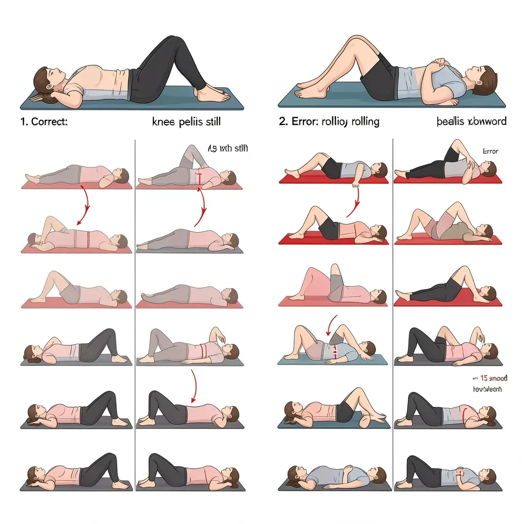
Clamshell
- Lie on your side with hips and knees at 45 degrees, pelvis stacked vertically
- Keep feet together and rotate the top knee upward, rotating from the hip
- Do NOT let the pelvis roll backward — this is the critical technique point
- The motion is small and should be felt in the lateral hip (gluteus medius), not the low back
- 15 repetitions each side, 2 sets
The clamshell builds gluteus medius endurance — the primary SIJ stabilizer during single-leg loading phases of gait.
Modified Bird-Dog with Hip Engagement
The standard bird-dog, modified to emphasize gluteal engagement at the start of each repetition:
- Start in the four-point kneeling position
- Before extending the leg, deliberately squeeze the gluteal muscles on that side (1-second hold)
- Then extend the leg into the bird-dog position while maintaining the squeeze
- The active hip extension with gluteal engagement activates the posterior SIJ stabilizers throughout the movement
- 10 repetitions each side, 2 sets
Symmetric Loading Pattern (Single-Leg Step-Up)
- Stand in front of a step or low box (15-20 cm)
- Brace the abdomen and actively engage the gluteal on the stepping leg
- Step up through the heel — not the toe
- Stand fully upright on the step with weight distributed evenly through the foot
- Step down with control
- The key: load the SIJ symmetrically through the foot-to-hip chain, not with a lateral shift or trunk lean
This exercise is a rehearsal for the stabilization pattern needed during every step of walking.
Aggravating Activities to Modify
- Long-stride walking on uneven terrain: Increases SIJ shear. Shorten stride and choose flat surfaces initially.
- Single-leg standing while dressing: Use a chair or wall for support when putting on pants, shoes, and socks.
- Car entry and exit: Getting into a car requires single-leg weight bearing while rotating the pelvis. Face away from the car seat, lower yourself onto the seat, then rotate both legs in together (same log-roll principle as getting out of bed).
- Crossing legs while seated: Unloads one SIJ and compresses the other chronically. Sit with feet flat and symmetrically placed.
- Carrying on one side: Bags, groceries, children — chronic unilateral loading. Distribute loads bilaterally or alternate sides.
- High-impact asymmetric sports: Running on cambered roads, throwing sports, single-leg dominant sport. Address SIJ stability before returning to these activities.
The most common rehabilitation error for SIJ dysfunction: prescribing hip flexor stretching and pelvic mobility work to "loosen" the area. The SIJ does not benefit from increased mobility. Aggressive hip flexor stretching can actually increase SIJ shear by pulling the ilium anteriorly relative to the sacrum. Focus entirely on stability, not flexibility, at this joint.
In Review
- The SIJ is a large, largely immobile joint that transfers all upper-body forces to the lower extremities; it is stabilized by massive ligaments and active muscular compression
- SIJ pain mimics L5-S1 disc herniation; pain below the knee is more consistent with true radiculopathy; focal pain over the PSIS is more consistent with SIJ
- The one-finger test (patient points precisely to the PSIS) is a useful screening differentiator
- Diagnosis requires multiple positive provocation tests (FABER, Gaenslen, thigh thrust, compression); three or more positive tests is highly specific
- Common causes: pregnancy, leg length discrepancy, asymmetric loading habits, post-lumbar fusion, trauma
- Mobilization of the SIJ is not therapeutic — the problem is instability, not restricted mobility
- The SI belt provides passive stabilization during demanding activities while muscular stability is being built
- The exercise focus: clamshell (gluteus medius endurance), modified bird-dog with hip engagement, symmetric loading patterns
- Modify asymmetric daily activities: distribute loads bilaterally, avoid single-leg standing, enter cars symmetrically