Lumbar Stenosis
Canal and foraminal stenosis — symptoms, why extension hurts, interval walking strategies, and long-term management.
What Stenosis Actually Is
Lumbar stenosis means narrowing of the spaces through which nerves travel in the lumbar spine. It is not a single entity but a structural consequence of the spine's normal aging cascade: disc height loss, facet joint enlargement from arthritis, ligamentum flavum thickening, and often the development of osteophytes (bone spurs).
The result is less space for nerves — and nerves under chronic compression or tension become sensitized, painful, and ultimately dysfunctional.
Canal stenosis: Narrowing of the central spinal canal, compressing the cauda equina (the bundle of nerve roots below the spinal cord). Produces bilateral or alternating symptoms, often affecting both legs.
Foraminal stenosis: Narrowing of the lateral recesses or intervertebral foramina — the exit holes through which individual nerve roots leave the spinal canal. Produces unilateral, dermatomal symptoms similar to disc herniation.
Both types commonly coexist, particularly in patients over 60.
Lumbar stenosis is the most common reason for spine surgery in adults over 65. It's also among the most overdiagnosed and oversurgicated conditions. Understanding the natural history — and what conservative management can accomplish — is essential context before any surgical conversation.
Neurogenic Claudication: The Signature Symptom Pattern
The characteristic symptom of lumbar canal stenosis is neurogenic claudication — a predictable pattern that distinguishes stenosis from nearly every other spine condition:
- Pain, heaviness, numbness, or weakness builds progressively in the legs with standing and walking
- Symptoms are relieved by sitting down or leaning forward (flexion)
- A specific walking distance triggers symptoms consistently
- The "distance to symptoms" may shorten over months or years as stenosis progresses
The walking distance that triggers symptoms is called the claudication distance. It is a clinically important measurement — track it over time.
The Shopping Cart Sign
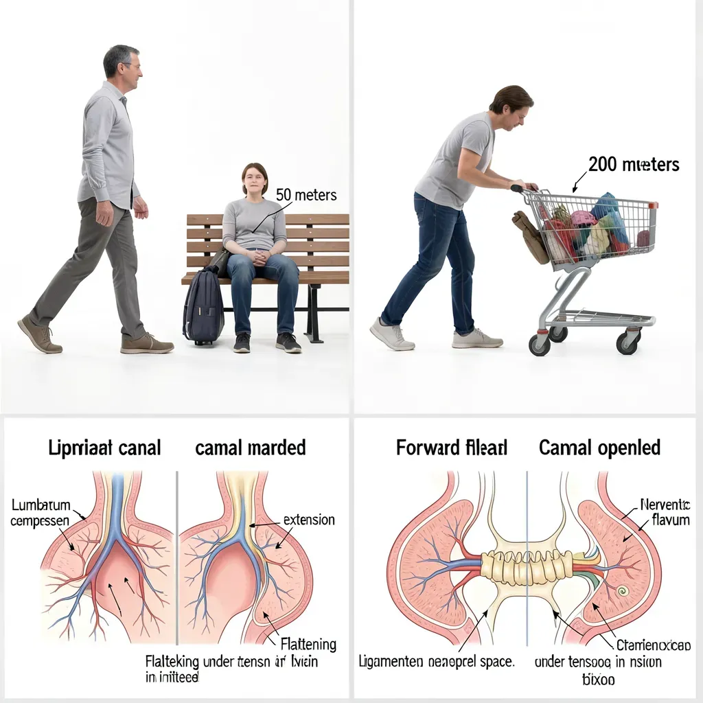
Stenosis patients instinctively lean forward over shopping carts, walkers, or countertops. This is not random. Lumbar flexion opens the spinal canal by stretching the ligamentum flavum taut and opening the facet joints posteriorly — gaining 15-30% more canal space in full flexion compared to full extension.
If a patient can walk substantially further pushing a shopping cart (which encourages a forward lean) than walking upright, neurogenic claudication is almost certainly the diagnosis.
Differentiating From Vascular Claudication
Both neurogenic and vascular claudication produce leg pain with walking that resolves with rest. The distinction matters because treatment is entirely different.
| Feature | Neurogenic Claudication | Vascular Claudication | |---------|------------------------|----------------------| | Relief position | Sitting or leaning forward | Standing still (any position) | | Relieved by stopping in place? | Usually no — must sit/flex | Yes — standing still relieves it | | Bicycle test | Can ride indefinitely (flexed posture) | Limited by same distance as walking | | Leg pulses | Normal | Reduced or absent | | Skin changes | Absent | Possible hair loss, skin thinning | | Age of onset | Typically 60s-70s | Typically 50s-60s with vascular risk factors |
The bicycle test is the most useful clinical differentiator. Cycling involves lumbar flexion throughout — it does not compress the stenotic canal. Neurogenic claudication patients can typically cycle much further than they can walk. Vascular claudication patients are equally limited on a bicycle.
If there is any question, ankle-brachial index testing (a simple vascular screening) rules out peripheral arterial disease quickly.
Why Extension Is Provocative
This is the central paradox of stenosis management — and the source of much misguided treatment:
The mechanism of extension pain: In lumbar extension, the ligamentum flavum buckles inward (it can't be placed under tension in extension), the facet joints close and approximate, and disc pressure redistributes posteriorly. The canal is at its narrowest in extension. For a canal with adequate space, this is inconsequential. For a stenotic canal, extension can directly compress neural tissue.
The management error: Many clinicians, observing that extension hurts and flexion helps, conclude that stenosis patients should stay in flexion. They prescribe flexion exercises and encourage a flexed posture.
This is a short-term relief / long-term harm trade.
Sustained lumbar flexion increases intradiscal pressure, compresses the disc, and over time accelerates the very degeneration that is creating the stenosis. Flexion provides neurological decompression but mechanical compression — simultaneously. The neurological benefit is immediate and noticeable; the mechanical harm is cumulative and silent.
Flexion relieves stenosis symptoms because it opens the canal momentarily. But sustained flexion posture compresses the discs, accelerates degeneration, and narrows the foramen over months and years. Use flexion as a rest-stop (a brief relief posture during a walk), not as a therapeutic posture. The goal is to build the capacity to walk in a neutral or near-neutral spinal position.
Interval Walking: The Primary Intervention
The most evidence-supported intervention for neurogenic claudication is a structured interval walking program. The principle: walk within your symptom-free window, rest briefly in a relief position, resume before symptoms return.
Protocol
-
Establish your claudication distance. Walk until you develop symptoms. Note the distance. This is your baseline.
-
Set your interval target at 50-75% of claudication distance. If you develop symptoms at 200 meters, your interval is 100-150 meters.
-
Walk the interval. Stop before symptoms appear. Lean against a wall in slight forward lean or sit for 60-90 seconds.
-
Resume walking when symptoms fully resolve. Do not push through.
-
Accumulate 20-30 minutes total walking time per session. Multiple short intervals count. The total volume is what matters for neural adaptation.
-
Progress the interval by 10% each week as your claudication distance improves.
This is not about building fitness in the traditional sense — though that also happens. It's about providing the repetitive compression-decompression cycle that promotes disc nutrition, maintains cartilage health, and prevents the vascular supply to the nerve roots from becoming chronically compromised.
Some stenosis patients find that a slight forward lean while walking (as if pushing slightly into a headwind) extends their walking distance dramatically. Experiment with trunk angle. A walking pole or trekking poles in both hands naturally encourages a slight forward lean and can double effective walking distance for many stenosis patients.
Decompression Exercises
These are positions and movements that actively open the spinal canal and provide temporary neural decompression. Use them at the end of intervals and as part of daily routine.
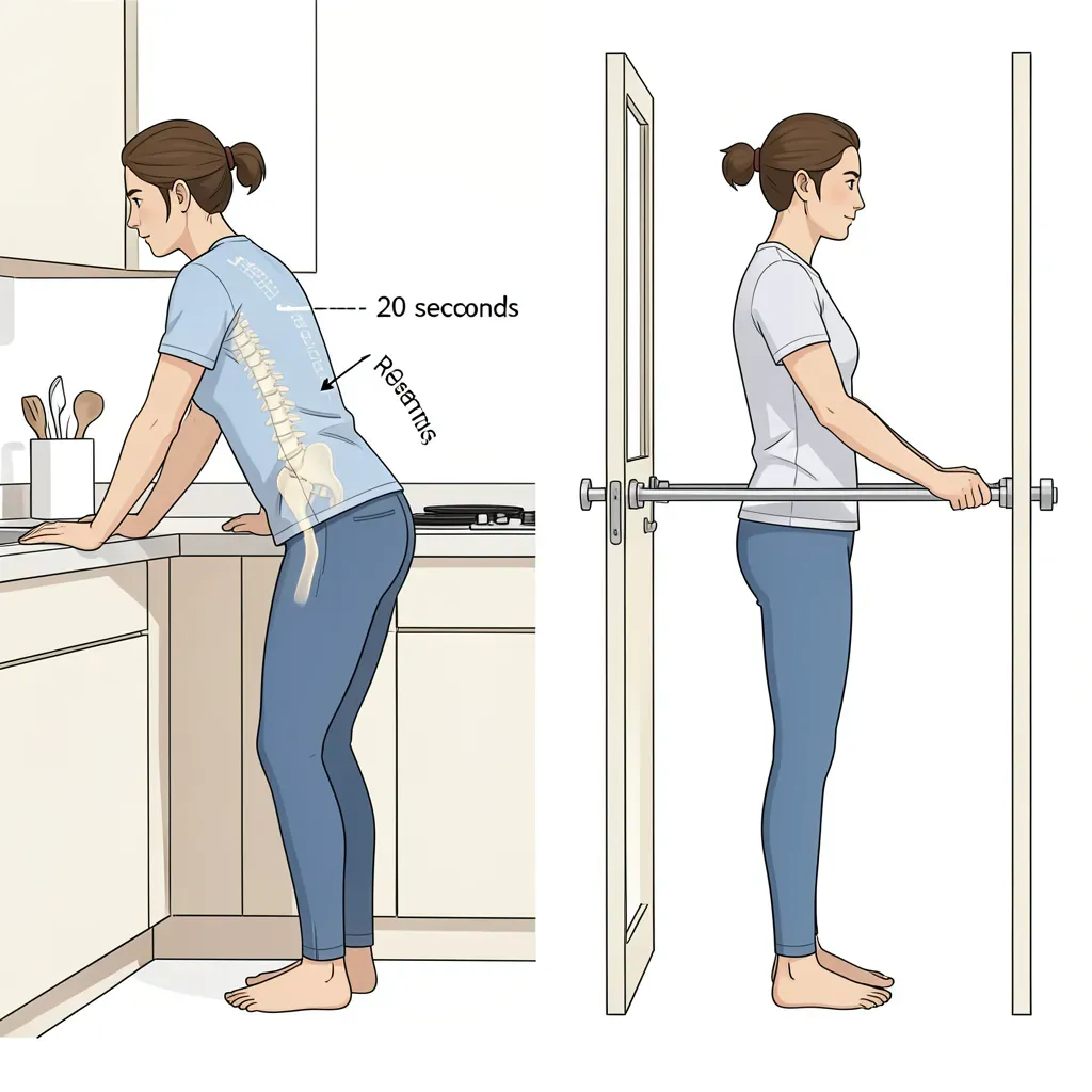
Standing hip hinge decompression:
- Stand facing a counter, railing, or table
- Step back 2-3 feet and grip the surface
- Hinge at the hips with knees soft, letting the spine lengthen
- Arms straight, slight traction through the spine
- Hold 20-30 seconds. The lumbar spine gently decompresses under its own weight with the hips as the hinge point.
Seated flexion stretch: Use sparingly — only as a symptom relief strategy, not a therapeutic exercise. Sit in a chair, lean forward onto your thighs. Hold 30 seconds. This relieves acute symptoms between walking intervals but is not a long-term therapeutic position.
Traction position: For more significant decompression: hang from a pull-up bar or doorframe chin-up bar with both hands, feet still touching the floor lightly. Allow gravity to provide gentle lumbar traction. 15-30 seconds. This is the most effective acute decompression position.
The Big 3 for Stenosis
The Big 3 exercises (curl-up, side bridge, bird-dog) are appropriate for stenosis patients with modifications:
Curl-up: Standard technique, Stage 1 and 2. The key is that the lumbar spine does not flex — the curl-up is an anti-flexion stability exercise despite its name. Appropriate and important for stenosis.
Side bridge: Standard from knees initially. Stenosis patients commonly have significant weakness in the lateral stabilizers from years of disuse. The side bridge rebuilds the lateral column stability that supports the spine without increasing axial load.
Bird-dog: The highest priority. The extended position of the bird-dog maintains a neutral-to-slight-extension spine while building posterior chain endurance. For stenosis, this is the exercise that builds tolerance for upright posture — the posture you need to improve walking distance.
Surgical Considerations
Conservative management should precede surgical discussion for any patient without progressive neurological deficit (worsening weakness or deteriorating bowel/bladder function).
When conservative management is appropriate (no deficit, symptoms manageable), outcomes at 2 years are similar between surgery and good conservative care in multiple randomized trials. Surgery provides faster relief; conservative care provides equivalent long-term outcomes with lower risk.
Surgical consultation is appropriate when:
- Progressive neurological deficit (weakness worsening over weeks)
- Bowel or bladder dysfunction
- Claudication distance has declined to the point where basic daily function is impossible
- Adequate trial of conservative management (minimum 3-6 months) has failed
The standard surgical procedure is decompression laminectomy — removing the bone and thickened ligament that is narrowing the canal. Outcomes are generally good for neurological symptoms. Pain relief is more variable.
Fusion is not required for most stenosis cases. Fusion is added when instability is present (as in spondylolisthesis — covered in the next article) or when extensive decompression would destabilize the joint. Most single or two-level decompressions do not require fusion. Be specific about what is being proposed and why.
Realistic Long-Term Outlook
Lumbar stenosis is a structural diagnosis that will not reverse. The goals of management are functional — maintaining walking capacity, quality of life, and independence.
With consistent interval walking and Big 3 maintenance:
- Claudication distance typically improves 30-50% over the first 3 months
- Many patients maintain functional walking capacity with ongoing management
- Symptoms fluctuate — more sedentary periods reliably worsen claudication distance; returning to active management reliably improves it
Stenosis is a condition you manage, not one you cure. The walking program is permanent, not a phase.
In Review
- Canal stenosis narrows the central spinal canal; foraminal stenosis narrows the nerve exit holes; both commonly coexist
- Neurogenic claudication is the signature pattern: walking triggers symptoms, sitting or leaning forward relieves them
- The shopping cart sign (extended walking with forward lean) is a reliable diagnostic indicator
- Distinguish from vascular claudication using the bicycle test and ankle-brachial index
- Extension provokes symptoms because the canal narrows; flexion relieves symptoms but causes long-term mechanical harm if used as a sustained posture
- Interval walking — walk within symptom-free distance, rest, repeat — is the primary intervention; progress intervals 10% weekly
- The Big 3 (especially bird-dog) build the stability needed for upright posture and extended walking
- Surgery is appropriate for progressive neurological deficit or failure of adequate conservative management; decompression laminectomy does not always require fusion