Reading Your MRI Report
What the terms in your MRI report actually mean — a plain-language guide to radiology language, common findings, and how to interpret them.
What You're Actually Reading
An MRI report is a radiologist's written description of what they observed in your scan. It follows a consistent structure: Clinical Indication (why the scan was ordered), Technique (what sequences were used), Findings (the detailed observations), and Impression (the radiologist's summary and conclusions). The Impression is what most doctors read first. It does not tell you what to do — it describes anatomy. That distinction matters enormously.
Radiologists are trained to document everything they see, including findings that have nothing to do with your symptoms. This is not careless; it's thorough. But it means reading a report without context can cause unnecessary panic.
Disc Findings: A Hierarchy
Not all disc problems are equal. The terminology follows a progression of severity:
A disc bulge is a broad, symmetric extension of disc material beyond the vertebral endplates — affecting more than 25% of the disc circumference. This is extremely common and often entirely asymptomatic. A disc protrusion is a focal herniation where the base is wider than the dome — the outer annular fibers are still intact. A disc extrusion means the nucleus has pushed through the annulus, with the extruded material still connected to the parent disc. A disc sequestration (or free fragment) means a piece of disc material has broken off entirely and migrated within the spinal canal.
Severity of your symptoms does not always match this hierarchy. Disc protrusions can cause severe radiculopathy; large bulges can cause nothing at all.
A 2014 study found that 52% of people with no back pain had at least one disc bulge on MRI. By age 60, that rises above 70%. Finding a disc bulge on your MRI does not automatically explain your pain.
Stenosis: Canal and Foraminal
Foraminal stenosis is narrowing of the opening through which a nerve root exits the spine. Reports typically grade it as mild, moderate, or severe. Mild foraminal stenosis is almost universally asymptomatic. Moderate stenosis may or may not cause nerve symptoms. Severe stenosis — particularly when accompanied by nerve compression on the scan — is more clinically significant, especially if your symptoms match the corresponding nerve's distribution.
Central canal stenosis is narrowing of the spinal canal itself. Reports sometimes cite the AP (anterior-posterior) diameter: below 10mm is considered absolute stenosis; 10–13mm is relative stenosis. Neurogenic claudication (leg pain and weakness with walking that improves with sitting or forward flexion) is the hallmark symptom.
Modic Changes and Endplate Findings
Modic changes describe signal alterations in the vertebral endplates — the bony surfaces above and below the disc. Type I changes show low signal on T1 and high signal on T2, indicating active inflammation and edema. This type correlates most strongly with pain and is considered the most clinically relevant. Type II shows high signal on both T1 and T2, indicating fatty replacement of the marrow — a stable, healed-over change, often asymptomatic. Type III shows low signal on both sequences and represents bony sclerosis. Types I and II can transition over time; Type I responding to treatment is a good prognostic sign.
Other Common Terms
Facet hypertrophy means the facet (zygapophyseal) joints have developed arthritic changes — bone remodeling and osteophyte formation. This is degenerative change and extremely common over age 50. Ligamentum flavum thickening describes hypertrophy of the ligament that lines the back of the spinal canal; it contributes to central stenosis when combined with disc bulging.
Spondylolisthesis is forward slippage of one vertebral body relative to the one below. MRI grading uses a percentage of the vertebral body width: Grade I is under 25%, Grade II is 25–50%. Most spondylolisthesis seen on MRI is degenerative (not traumatic) and Grade I.
The phrase "multilevel degenerative changes" on an MRI report sounds alarming but is nearly universal in adults over 40. It describes the normal aging process of spinal structures — not necessarily a cause of pain, and not a predictor of future disability.
The Incidentaloma Problem
Incidentalomas are findings discovered incidentally — things visible on your scan that were not causing symptoms and were not the reason the scan was ordered. MRI is so sensitive that it routinely detects cysts, small hemangiomas, minor bone marrow changes, and structural variants that are clinically irrelevant. The challenge is distinguishing findings that explain your pain from findings that are passengers. This requires clinical correlation — matching the imaging to your symptoms, physical examination findings, and history. No radiologist can do this from the scanner room. Your clinician must make that judgment.
Discussing Your MRI with Your Doctor
Come prepared. Write down your specific symptoms: which leg, what type of sensation (burning, shooting, numbness), what aggravates and relieves it. Ask your doctor which finding on the report they believe is responsible for your symptoms, and why. Ask whether the findings match your symptoms clinically. A good clinician will not treat the report — they will treat you.
Critical Findings That Require Urgent Attention
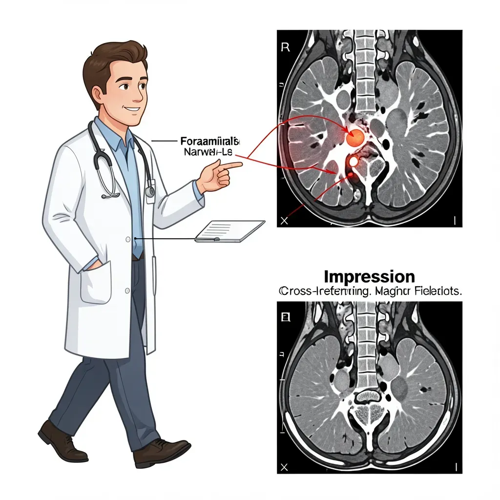
Most MRI findings are not emergencies. These are: spinal cord compression with signal change in the cord (myelopathy on T2 signal) — this requires urgent surgical evaluation. Cauda equina syndrome — compression of the cauda equina nerve roots causing loss of bowel or bladder control, saddle anesthesia, or bilateral leg weakness — is a surgical emergency. If your MRI is accompanied by these symptoms, go to the emergency department. Do not wait for a follow-up appointment.
In Review
- MRI reports describe anatomy, not your destiny — findings must be correlated with your symptoms
- Disc bulges and "multilevel degenerative changes" are common in pain-free adults; their presence on your scan doesn't automatically explain your pain
- Disc severity follows a hierarchy: bulge, protrusion, extrusion, sequestration — but symptoms don't always match the imaging severity
- Modic Type I changes are the most clinically significant endplate finding; Types II and III are often incidental
- Mild and moderate foraminal stenosis is frequently asymptomatic; severe stenosis warrants closer clinical correlation
- Cauda equina syndrome is the one MRI finding that constitutes a true emergency
- Always ask your doctor which specific finding they believe is causing your symptoms, and why