Facet Arthritis & Bone Spurs
How osteoarthritis develops in the facet joints, what bone spurs mean clinically, and a management approach for facet-dominant pain.
What Are the Facet Joints?
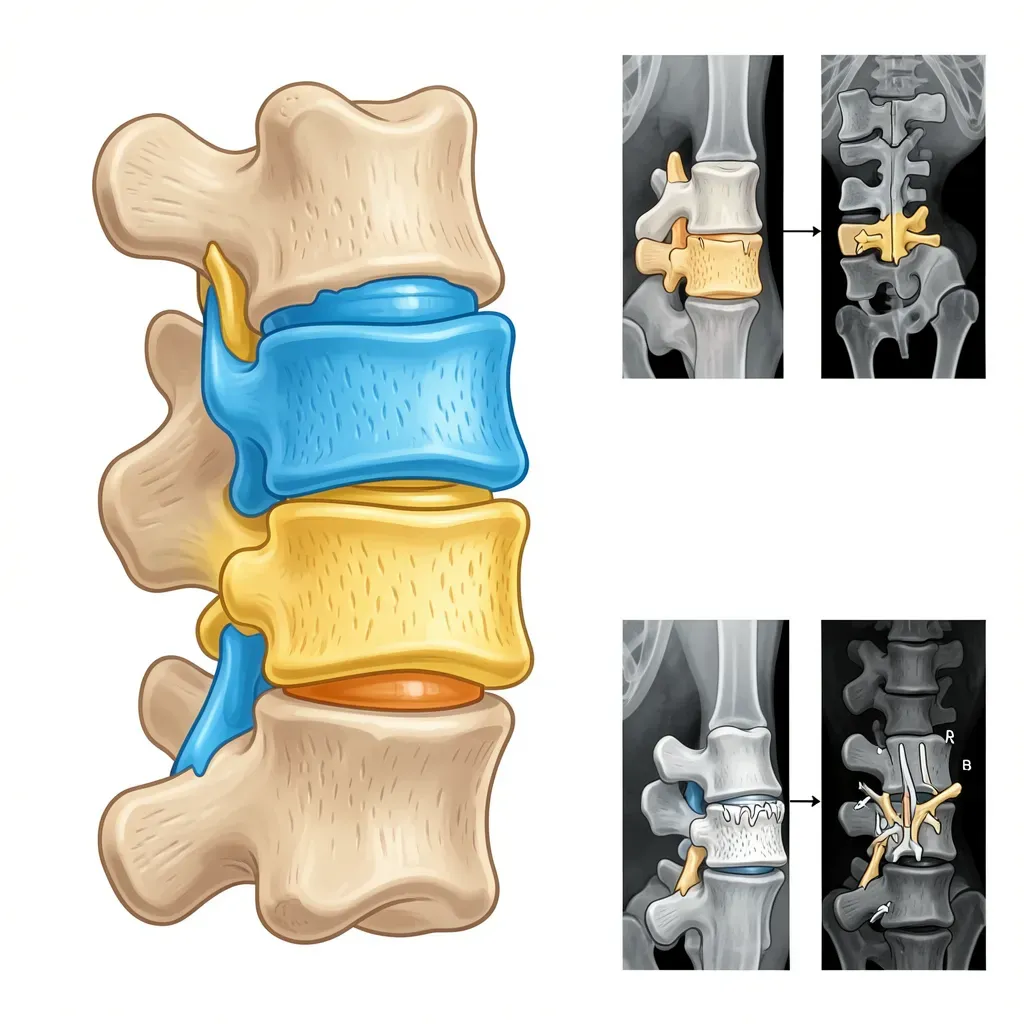
Each vertebral level has two facet joints (also called zygapophyseal joints, or Z-joints) — one on each side. They are true synovial joints: cartilage-lined, encapsulated, lubricated with synovial fluid, and innervated by the medial branches of the dorsal rami (two nerves per joint, from the level above and below).
Their orientation determines what motion each spinal region allows. In the lumbar spine, the facets are oriented roughly in the sagittal plane, allowing flexion and extension but resisting rotation. In the thoracic spine, they're more coronally oriented, allowing rotation. The cervical facets allow the most motion in all planes.
Functionally, the facet joints share compressive load with the disc. A healthy disc carries the majority of compressive force. As a disc degenerates and loses height, the facet joints on the same segment absorb more load — transitioning from secondary to primary load-bearers. They are not designed for this role. This load transfer is the primary initiating event for facet osteoarthritis.
The OA Process in Facet Joints
Osteoarthritis in the facet joints follows the same pathological sequence as OA in any synovial joint:
Cartilage degradation: The articular cartilage, which has no blood supply and limited regenerative capacity, begins to soften and fibrillate. The smooth, low-friction surface becomes irregular and rough.
Subchondral bone changes: The bone beneath the cartilage thickens (sclerosis) in response to increased stress. This is the "white on MRI" appearance on T1-weighted images around arthritic joints.
Joint space narrowing: As cartilage is lost, the space between the articulating surfaces decreases. Bone-on-bone contact occurs in advanced stages.
Synovitis: The synovial lining becomes inflamed in active OA stages, producing the morning stiffness and activity-related flares that characterize the condition.
Osteophyte formation: New bone grows at the joint margins — an attempted stabilization response. The spine is trying to fuse the unstable, painful segment.
Facet osteoarthritis is not simply "wear and tear" from overuse. It is primarily driven by abnormal load distribution — usually from disc degeneration transferring load to structures not built for it. This means that managing load distribution (disc health, posture, muscle function) is the genuine upstream intervention, not just managing the facet joint symptoms downstream.
The Clinical Presentation
Facet-dominant pain has a recognizable pattern that helps distinguish it from disc-dominant pain:
Provocative directions: Extension and rotation are typically the aggravating movements. This is the opposite of disc pain, which is usually aggravated by flexion. A patient who feels worse arching backward, whose symptoms return at the end of a long walk, or who cannot comfortably stand in extension — facet pathology is high on the differential.
Morning stiffness: The synovitis component of facet OA produces classic joint stiffness that improves after 10-15 minutes of movement. This is distinct from disc pain, which often eases with lying down and worsens with sustained positions.
Activity pattern: Facet pain typically worsens throughout the day with activity, unlike disc pain which is often worst in the morning before the nucleus rehydrates. Standing for prolonged periods and walking downhill (which increases lumbar extension) are common aggravators.
Pain location: Central or paraspinal lower back pain, often with a deep aching quality. Referred pain can extend into the buttock and posterior thigh — but rarely below the knee, which distinguishes it from true radiculopathy. This referred pain pattern from facet capsule irritation is called "somatic referred pain" — it follows non-dermatomal distributions and does not have the burning, electric quality of nerve root compression.
Palpation: Firm palpation at the paraspinal muscles at the affected levels typically reproduces the patient's pain. Unlike disc herniation, where palpation may be unremarkable, facet arthritis produces consistent local tenderness.
The clinical pattern of facet pain — worse with extension and rotation — means that common rehabilitation advice to "arch your back" or "do back extensions" can directly aggravate facet-dominant presentations. Extension exercises are appropriate for disc herniation but contraindicated as a primary intervention for facet arthritis. Identifying which structure is driving the pain before prescribing directional exercises is not optional.
Osteophytes: Stabilization, Not Just Damage
Bone spurs (osteophytes) routinely alarm patients who see them on imaging reports. The framing of "bone spurs" as sharp objects stabbing soft tissue is common in lay understanding but largely inaccurate.
Osteophytes are smooth, rounded bony outgrowths at joint margins. They form as a response to instability — the periosteum (bone surface) is stimulated by abnormal stress to deposit new bone, incrementally bridging the unstable joint. In advanced degeneration, adjacent osteophytes may actually fuse across the disc space, creating a spontaneous fusion — the pain sometimes resolving when this stabilization is complete.
The clinical reality: some of the most dramatically osteophyte-laden spines on X-ray belong to people with minimal or no symptoms. Large osteophytes are often a sign that the stabilization process has already largely completed. Conversely, a spine with minimal osteophyte formation but significant disc height loss may still be in the painful, unstable intermediate phase.
Osteophytes can cause problems through two mechanisms: nerve root encroachment in the foramina (producing radiculopathy) or spinal canal encroachment (contributing to stenosis). These specific complications require specific assessment — but the osteophyte itself is not the problem in most cases.
Modifiable Factors
Two modifiable factors have direct relevance to facet joint load:
Body weight: Each kilogram of body weight above ideal increases compressive force on the lumbar spine by roughly 4-8 kilograms at L4-L5 (depending on lever arm calculations and activity). Adiposity also has an inflammatory component — adipose tissue is metabolically active and produces pro-inflammatory cytokines that worsen joint inflammation systemically. Weight loss, where applicable, is one of the highest-impact interventions available for facet OA.
Abdominal and hip musculature: The facet joints share compressive load with the disc and the surrounding musculature. Strong paraspinal and abdominal muscles that can generate intra-abdominal pressure during loading distribute compressive forces across more structures — reducing the share borne by any single component. This is the mechanical rationale for core endurance training in facet OA management.
Exercise Approach
The central principle for facet-dominant pain: train in neutral, not into extension.
Appropriate: McGill Big 3 (curl-up, side plank, bird-dog) in neutral spine, glute strengthening (bridges, clamshells), hip flexor stretching (which reduces anterior pelvic tilt and lumbar extension moment), walking on level surfaces, swimming (especially freestyle, which keeps the spine relatively neutral).
Modify or avoid: Deep lumbar extension exercises, standing back bends, loaded rotation, walking on inclines, heavy overhead pressing (which compresses the facets in extension under load).
As symptoms stabilize, the program can progressively increase load and variety. The goal is not permanent extension avoidance — it is appropriate load management during the symptomatic phase, followed by rebuilding robust load tolerance.
Facet Injections: Diagnostic and Therapeutic
Medial branch blocks (local anesthetic injected around the nerve supply to the facet joint) are both diagnostic and therapeutic. If the block produces complete, temporary pain relief — confirming the facet joint as the pain source — and the effect wears off, radiofrequency ablation (burning the medial branch nerves) can provide 6-24 months of pain relief.
This is not a cure — the nerves regenerate — but it can provide a meaningful pain-free window to complete rehabilitation that would otherwise be too painful to pursue.
Facet joint injections should be combined with rehabilitation, not used as a stand-alone treatment. The injection reduces pain to allow exercise; the exercise rebuilds the muscular support that reduces load on the joint long-term. Repeated injections without rehabilitation typically produce diminishing returns.
Long-Term Prognosis
Facet OA follows the general OA prognosis: gradual but rarely catastrophic progression, with substantial variability based on load management, body weight, and physical fitness. Many patients reach a stable plateau where symptoms are manageable and do not progress significantly over years. Severe cases — particularly those with significant foraminal stenosis from osteophyte encroachment — may require surgical intervention (foraminotomy), but this is a minority of presentations.
The key clinical message: facet arthritis is a manageable condition. It is not a death sentence for an active life, and the majority of people who approach it with appropriate load management and consistent exercise maintain good quality of life.
In Review
- Facet joints are synovial joints that bear compressive load alongside the disc; disc degeneration shifts load to facets, initiating OA
- Clinical pattern: worse with extension and rotation, morning stiffness, worsens through the day — the opposite of disc-dominant pain
- Osteophytes are a stabilization response, not random damage — large osteophytes often indicate near-completed stabilization
- Exercise in neutral spine, not into extension; hip flexor stretching and gluteal strengthening are core components
- Body weight and core strength are the primary modifiable variables for facet load
- Medial branch blocks confirm diagnosis; radiofrequency ablation provides extended relief; should always be combined with rehabilitation