Ankylosing Spondylitis: Exercise and Spine Management
The unique challenges of ankylosing spondylitis — why the standard back pain advice doesn't apply, how fusion affects mechanics, and the specific exercise priorities.
A Fundamentally Different Disease
Ankylosing spondylitis (AS) is an inflammatory arthritis of the spine and sacroiliac joints. It is not a musculoskeletal overuse injury, a disc problem, or a postural issue. It is an immune-mediated disease in which the body's inflammatory response targets the entheses — the points where ligaments, tendons, and joint capsules attach to bone — throughout the axial skeleton. Over time, this inflammatory process triggers bone formation at those attachment sites, progressively bridging vertebral segments and eventually fusing the spine.
This distinction has profound implications for management. Most of what is known about back pain — rest when it hurts, protect the spine from load, avoid positions that provoke pain — does not apply to ankylosing spondylitis. In fact, applying those principles to AS reliably produces worse outcomes. Understanding why requires understanding the disease's unique mechanics.
The Genetics and Early Presentation
Approximately 90–95% of people with AS carry the HLA-B27 genetic marker, though only a small fraction of HLA-B27-positive individuals develop the disease. AS most commonly presents in the late teens to mid-thirties, with an insidious onset of low back and buttock pain that is characteristically worse in the morning and with inactivity, and better with movement and exercise. This inflammatory back pain pattern — morning stiffness lasting more than 45 minutes, improvement with movement, and no improvement with rest — is the clinical fingerprint that distinguishes AS from mechanical back pain.
The sacroiliac joints are almost always affected first. Inflammation at the SI joint produces pain in the buttock, posterior hip, and sometimes the posterior thigh that can be mistaken for piriformis syndrome or lumbar radiculopathy. As the disease progresses, the inflammatory front moves up the spine through the lumbar, thoracic, and eventually cervical vertebrae.
The average time from symptom onset to AS diagnosis is historically 7–10 years. This delay occurs because AS in young adults is often attributed to mechanical back pain, muscle strains, or sports injuries. Young men (AS is two to three times more common in men) with insidious-onset inflammatory back pain should be evaluated for AS, particularly if there is a family history of the disease or associated features such as uveitis, psoriasis, or inflammatory bowel disease.
Why Rest Worsens AS
In mechanical back pain, rest is often temporarily beneficial — it reduces load on an irritated structure and allows acute inflammation or tissue disruption to subside. In AS, rest is counterproductive at every stage of the disease.
The reason is both biological and biomechanical. Biologically, the inflammatory process in AS continues regardless of whether the patient is active or sedentary. There is no period of rest that reduces the underlying immune-mediated inflammation. The pain that comes from inactivity in AS reflects the accumulation of inflammatory mediators in stiff, poorly perfused joint tissues during periods of immobility — which is why morning stiffness is the dominant symptom.
Biomechanically, as the disease progresses and the spine begins to fuse, the position in which it fuses becomes critically important. A spine that fuses in a kyphotic posture — which happens when patients protect it by avoiding extension, sitting and sleeping in flexed positions, and avoiding exercise — will leave the patient permanently stooped, with cervical hyperextension as the only way to level the gaze. A spine that fuses in a more neutral, upright position — which requires aggressive exercise and postural maintenance throughout the disease course — preserves far more function.
This is the most important clinical point about AS: exercise is not just beneficial. It is a primary disease-management strategy. The evidence clearly shows that people with AS who exercise consistently have better spinal mobility, less functional disability, and slower functional decline compared to those who do not.
Sacroiliac Joint Inflammation
The sacroiliac joint in AS is a true synovial joint in its anterior ligamentous portion and an amphiarthrodial joint (cartilaginous) in its posterior portion. Inflammatory involvement of the SI joint in AS causes sacroiliitis — detectable on MRI as bone marrow edema before structural damage is visible on X-ray — which is the earliest structural sign of the disease.
The pain from sacroiliitis is often bilateral or alternating, deep in the buttock, and worse after prolonged sitting or sleeping. It tends to improve after 30–60 minutes of morning movement. SI joint provocation tests (posterior pelvic pressure, FABER, FADIR) may be positive but are not specific to AS sacroiliitis. MRI of the SI joints is the most sensitive imaging modality for early disease.
As sacroiliitis progresses, the SI joints fuse. Once fused, the pain from the SI joints typically resolves — the joint is no longer moving, so it can no longer be irritated by movement. This is a counterintuitive feature of AS: some patients feel better as their disease advances, because the pain source fuses. However, the functional consequences of fusion — reduced spinal mobility, altered gait mechanics, vulnerability to fracture — are the long-term challenges that exercise addresses.
Progressive Fusion: Mechanical Consequences
As AS progresses and spinal segments fuse, the mechanical behavior of the spine changes fundamentally. A partially or completely fused spine does not move the way an intact spine moves. Load that would normally be distributed across many motion segments is instead concentrated at the junctions between fused and unfused segments, or at the ends of a long fused column (the cervicothoracic and lumbosacral junctions).
The fused spine is also brittle in a way that a healthy spine is not. Because the vertebrae are bridged by bone rather than connected by flexible ligaments and disc material, the entire fused column behaves like a long bone. A fall or unexpected impact that a normal spine would absorb through multi-segmental load distribution can fracture through a fused spine — these are called pseudo-arthrosis fractures and can occur at any level, including with relatively minor trauma. People with long-standing AS who develop acute severe spinal pain after any kind of mechanical event require immediate imaging.
People with advanced ankylosing spondylitis who sustain any fall, collision, or unexpected mechanical load to the spine should seek medical evaluation before resuming exercise. The fused spine is vulnerable to fracture from mechanisms that would be trivial in a healthy spine, and these fractures can threaten the spinal cord. Never assume spine pain after a physical event is "just the AS."
Swimming and Breathing
Swimming is consistently highlighted as one of the best exercise modalities for AS patients, and the reasons are biomechanically specific. In the water, the spine is unloaded from compression, allowing movement that would be painful under axial load on land. Swimming backstroke and freestyle require thoracic extension and rotation, which are the mobility qualities that AS specifically threatens. The rhythmic, repetitive movements of swimming maintain joint and soft tissue mobility throughout the spinal column.
Breathing exercises have a unique importance in AS. The thoracic spine's attachment to the rib cage means that thoracic fusion directly compromises chest wall expansion. Reduced chest expansion — measured as the difference in chest circumference between full expiration and full inspiration — is a formal clinical measure used to track AS progression. As the thoracic spine and costovertebral joints fuse, chest expansion is restricted, forcing the diaphragm to do a larger share of respiratory work and reducing overall respiratory reserve.
Deep breathing exercises, which actively expand the chest wall and costovertebral joints, should be incorporated daily. Simple exercises: maximal inspiration held for 5 seconds, lateral chest expansion with hands placed on the sides of the chest, and prone extension with deep breathing all combine mobility maintenance with respiratory training.
Thoracic Mobility: The Priority
Of all the regions of the spine affected by AS, the thoracic spine deserves particular exercise priority for three reasons. First, it fuses earliest and most completely in many patients. Second, thoracic fusion has the greatest impact on quality of life — it drives the characteristic stooped posture, limits chest expansion, restricts overhead reach, and determines whether the patient can see where they are walking. Third, thoracic mobility is the most amenable to exercise intervention in early-to-moderate disease.
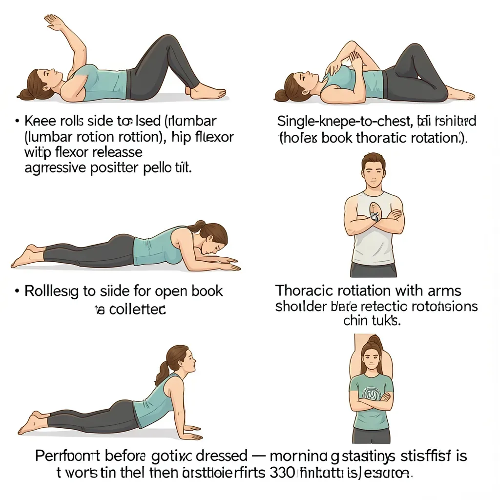
Exercises for thoracic mobility in AS: foam roller extension, open book rotations, prone extension (cobra-type movements), thoracic rotation in quadruped, and specific NASS (National Ankylosing Spondylitis Society) protocols that combine extension and rotation. These should be performed daily, not occasionally. The morning stiffness of AS means that the first mobility exercises of the day are both the hardest and the most important — beginning the day with 10–15 minutes of thoracic and hip mobility work substantially reduces morning stiffness duration.
NSAIDs, Biologics, and Exercise
Nonsteroidal anti-inflammatory drugs (NSAIDs) — particularly indomethacin, naproxen, and diclofenac — are the first-line medical treatment for AS and have a unique role that extends beyond pain relief. There is evidence that continuous NSAID use may slow the rate of radiographic progression (bony fusion) in AS, possibly because the inflammatory process that drives fusion is partly inhibited by NSAID-mediated COX pathway suppression. This makes NSAID use a disease-modifying strategy in AS, not just a symptomatic one.
Biological disease-modifying antirheumatic drugs (bDMARDs) — tumor necrosis factor inhibitors (adalimumab, etanercept, infliximab) and IL-17 inhibitors (secukinumab, ixekizumab) — have transformed outcomes for AS patients over the past two decades. They dramatically reduce inflammation, improve symptoms and function, and appear to slow structural damage. For patients whose disease is not controlled by NSAIDs, bDMARDs are the next treatment step.
The relevance to exercise is direct. Patients on effective biological therapy often experience substantial reduction in pain and stiffness, which dramatically improves their ability to exercise. The combination of biologic therapy and structured exercise produces better outcomes than either alone. This is not an argument for delaying exercise until medication is optimized — exercise should begin as early as possible — but it is an argument for ensuring that patients with active, poorly controlled disease have access to appropriate medical treatment so that exercise can be performed effectively.
Posture Maintenance as Disease Management
In AS, posture is not cosmetic — it is functional disease management. The position in which the spine fuses determines the patient's lifelong functional capacity. Patients who consistently maintain extension-biased posture, sleep on firm flat surfaces without thick pillows, avoid prolonged sitting in flexed postures, and perform daily extension exercises preserve spinal alignment that translates directly to better lifelong function.
Sleeping position matters particularly. Sleeping prone (face down) or supine with a flat pillow maintains spinal extension. Sleeping in the fetal position — curled on the side with hips and knees bent, thoracic spine rounded forward — is the position AS will fix the spine in if given the chance.
In Review
- Ankylosing spondylitis is an immune-mediated inflammatory disease of spinal entheses, not a mechanical injury — the standard back pain advice of rest and protection is actively harmful
- The cardinal symptom pattern — morning stiffness lasting more than 45 minutes, worsening with rest, improving with movement — distinguishes AS from mechanical back pain
- HLA-B27 positivity is present in 90–95% of AS patients; early sacroiliitis is best detected on MRI before X-ray changes appear
- Progressive spinal fusion makes posture maintenance a life-altering priority; the spine will fuse in the position it is most frequently held
- A fused spine behaves like a long bone and is vulnerable to fracture from impacts that would be trivial in a healthy spine — any mechanical event causing acute spinal pain requires imaging
- Swimming and daily thoracic mobility exercises are the most important specific exercise interventions; chest expansion exercises address the unique respiratory consequences of thoracic fusion
- NSAIDs have disease-modifying potential in AS beyond symptom relief; biologic agents markedly improve disease control and enable more effective exercise
- Exercise is not an adjunct in AS management — it is a primary intervention with effects on functional trajectory that are as important as medical therapy