Back Pain and Obesity: Mechanisms Beyond Simple Load
Why obesity is more than just extra weight on the spine — adipose tissue as an endocrine organ, the inflammatory mediators produced, and how weight loss affects pain.
Reframing the Obesity-Back Pain Relationship
The conventional explanation of obesity's contribution to back pain is mechanical: more weight means more load on the spine. This explanation is not wrong — it is just severely incomplete. It treats adipose tissue as inert ballast when in fact it is a metabolically active endocrine organ, continuously secreting hormones and cytokines that directly influence spinal inflammation, pain processing, and tissue health. And it fails to explain several clinical observations that the simple mechanical model cannot account for: why upper-body obesity is more strongly associated with back pain than lower-body obesity, why weight loss produces pain improvements disproportionate to the mechanical unloading alone, and why interventions that reduce systemic inflammation improve back pain even without significant weight change.
A more complete model treats obesity as simultaneously a mechanical, hormonal, and inflammatory condition — and recognizes that these three mechanisms are additive and interacting, not independent. Managing the obesity-back pain relationship requires addressing all three.
Adipose Tissue as an Endocrine Organ
Until the 1990s, adipose tissue was understood primarily as energy storage. The discovery of leptin in 1994 — a hormone produced by fat cells — fundamentally changed this understanding. Adipose tissue is now recognized as one of the most metabolically active tissues in the body, secreting over 600 signaling proteins collectively called adipokines. These molecules regulate appetite, insulin sensitivity, immune function, angiogenesis, and inflammation.
In the context of back pain, three adipokines are most clinically relevant:
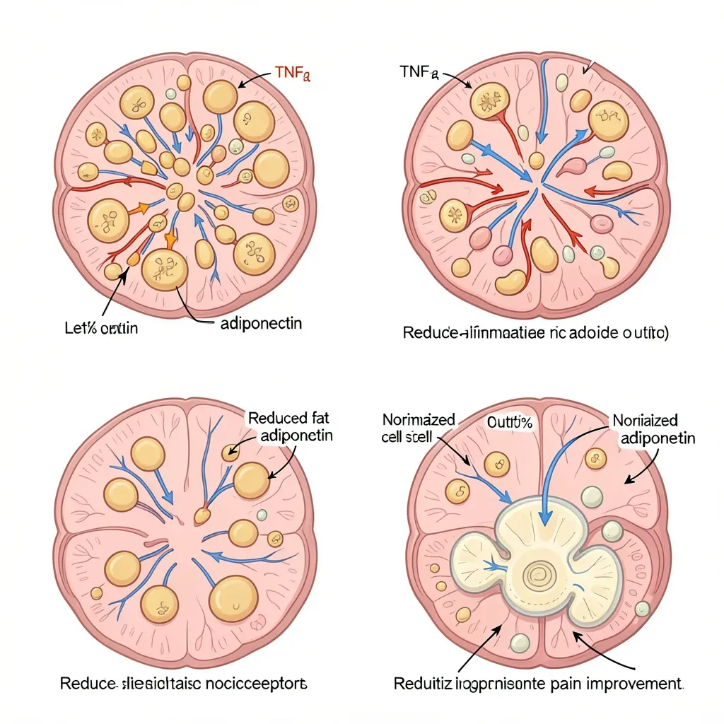
Leptin — produced in proportion to fat mass; regulates appetite and metabolism via hypothalamic receptors, but also acts on immune cells and joint tissue. In obese states, leptin levels are chronically elevated. Leptin promotes the production of pro-inflammatory cytokines (IL-6, TNF-α, IL-12) and directly stimulates joint and disc nociceptors. Leptin receptors are present in intervertebral disc tissue, and in vitro studies show that leptin exposure promotes disc cell catabolism — the breakdown of disc matrix. Higher serum leptin is independently associated with disc degeneration severity in imaging studies, even after controlling for BMI.
Adiponectin — unlike most adipokines, adiponectin has anti-inflammatory effects. It inhibits NF-κB signaling (the master regulator of inflammatory cytokine production), promotes insulin sensitivity, and has protective effects on articular cartilage. Critically, adiponectin levels are inversely proportional to fat mass — obese individuals have lower adiponectin than lean individuals. As obesity progresses, the ratio of pro-inflammatory leptin to anti-inflammatory adiponectin shifts unfavorably: more inflammation-driving signal, less inflammation-suppressing signal.
TNF-α from adipose tissue — while TNF-α (tumor necrosis factor-alpha) is produced by multiple immune cell types, visceral adipose tissue is a significant source. TNF-α directly sensitizes nociceptors in disc and joint tissue, promotes matrix metalloproteinase (MMP) production that degrades disc matrix, and drives the systemic inflammatory state that amplifies pain at the central level.
Visceral vs. Subcutaneous Fat: Different Inflammatory Profiles
Not all adipose tissue is equivalent. Visceral adipose tissue (VAT) — the fat deposited within the abdominal cavity, surrounding the organs — has a fundamentally different inflammatory profile from subcutaneous adipose tissue (SAT), the fat stored under the skin.
Visceral adipose tissue:
- Has higher macrophage infiltration, particularly inflammatory M1-polarized macrophages
- Produces more TNF-α, IL-6, and leptin per unit mass than subcutaneous fat
- Drains directly into the portal circulation, exposing the liver to high concentrations of inflammatory cytokines and free fatty acids — driving hepatic insulin resistance and further systemic inflammation
- Has higher lipolytic activity, generating more circulating free fatty acids that activate inflammatory signaling (toll-like receptor 4 activation)
Subcutaneous adipose tissue, particularly gluteal and femoral fat, is metabolically relatively quiescent — it stores and releases fatty acids but produces fewer inflammatory cytokines and is not directly associated with the metabolic and inflammatory complications of obesity.
This distinction explains the clinical observation that waist circumference and waist-to-hip ratio predict health outcomes and pain burden better than BMI alone. Two people with identical BMI can have dramatically different visceral fat content depending on fat distribution pattern — and the one with higher visceral fat will have a more inflammatory metabolic profile. For back pain specifically, waist circumference above 102 cm in men and 88 cm in women (the ATP III criteria for abdominal obesity) is a more meaningful risk indicator than BMI category alone.
Mechanical Effects: Anterior Pelvic Tilt and Spinal Load
The mechanical contribution is real and clinically relevant — it is simply not the complete story. Excess abdominal load shifts the center of gravity anterior to the spine, creating an increased moment arm that requires compensatory hyperextension of the lumbar spine and greater paraspinal muscle activation to maintain upright posture. The result is a predictable pattern: increased lumbar lordosis (anterior pelvic tilt from abdominal load), chronically increased paraspinal muscle tension, and elevated compressive load on the facet joints and posterior disc annulus.
Biomechanical models estimate that each pound of abdominal fat increases the compressive load on the lumbar spine by approximately 4 pounds through this moment arm amplification. For someone carrying 40 excess pounds of central adiposity, this translates to roughly 160 extra pounds of lumbar compressive load in upright posture — a meaningful structural burden that explains why obesity is such a strong predictor of disc degeneration and facet arthritis progression on imaging.
The effect on disc hydration is also relevant: sustained high compressive loads (as described in chapter 114 for athletes) exceed the disc's capacity to maintain fluid balance. The chronically elevated lumbar compressive load from obesity is a constant mechanical stressor that the disc cannot adequately recover from overnight in the way it might with normal-weight loading.
Why 10% Weight Loss Produces Disproportionate Pain Improvement
Clinical observation, reinforced by several studies, shows that a 10% reduction in body weight produces pain improvements that exceed what the mechanical unloading alone would predict. A 10% weight reduction in a 120 kg patient — 12 kg — does not produce a mechanically major change in spinal compressive load. Yet functional outcomes and pain scores often improve substantially.
The disproportionate improvement is explained by the non-linear relationship between fat mass and inflammatory mediator production. Adipose tissue reaches an inflammatory tipping point as fat cell size increases. Enlarged adipocytes (hypertrophic fat cells in obesity) are metabolically stressed, hypoxic, and infiltrated with inflammatory macrophages. As these cells shrink with weight loss, their inflammatory output decreases non-linearly — a relatively small reduction in fat cell size produces a large reduction in inflammatory cytokine production.
Additionally, 10% weight loss is associated with measurable improvements in adiponectin levels (which were suppressed), further shifting the leptin:adiponectin ratio toward the anti-inflammatory end. The systemic inflammatory environment — reflected in CRP, IL-6, and TNF-α levels — improves significantly with 10% weight loss in obese individuals, even when absolute BMI remains in the obese range.
The clinical implication is important for goal-setting with obese back pain patients: the threshold for meaningful benefit is not "getting to a healthy BMI" — it is achieving 5–10% weight reduction. This is a psychologically important reframe for patients who feel that the distance from their current weight to a "healthy" BMI is so large as to be irrelevant to back pain. It is not.
Exercise vs. Diet for Pain Specifically
When weight loss is the goal, both diet-driven caloric restriction and exercise-driven caloric expenditure can achieve similar weight loss results over time. However, for back pain specifically, the mechanism of weight loss matters.
Exercise produces benefits for back pain through multiple pathways that operate independently of any weight change:
- Direct anti-inflammatory effects of aerobic exercise (IL-6 produced by contracting muscle has anti-inflammatory actions distinct from TNF-α-derived IL-6; regular exercise reduces resting CRP, TNF-α, and IL-6 levels)
- Paraspinal and core muscle strengthening that reduces mechanical load concentration
- Improved disc nutrition through cyclic loading
- Central sensitization reduction through endogenous opioid and endocannabinoid system activation during exercise
- Improved sleep quality, which reduces pain threshold impairment
Diet-driven weight loss achieves the metabolic improvements (reduced leptin, increased adiponectin, reduced visceral fat inflammatory output) but does not produce the direct structural and neurological benefits that exercise provides. The combination — exercise plus modest caloric deficit — is clearly superior to either alone for back pain outcomes.
For back pain patients who are obese and significantly deconditioned, the practical starting point is always low-impact activity that does not aggravate their pain: walking, swimming, aquatic exercise, recumbent cycling. The goal is progressive daily movement rather than a formalized exercise program, initially. As pain and capacity improve, structured resistance training can be added — and the resistance training component has the additional benefit of increasing resting metabolic rate and preserving muscle mass during caloric restriction.
In Review
- Adipose tissue is an endocrine organ producing adipokines — including leptin, adiponectin, and TNF-α — that directly influence spinal disc health and pain processing
- Leptin (elevated in obesity) promotes pro-inflammatory cytokine production and directly stimulates disc nociceptors; adiponectin (reduced in obesity) has anti-inflammatory protective effects
- Visceral fat is substantially more inflammatory than subcutaneous fat — waist circumference is a more meaningful predictor of back pain burden than BMI alone
- Each pound of central abdominal fat adds approximately 4 pounds of lumbar compressive load through moment arm amplification, producing anterior pelvic tilt and facet joint overloading
- A 10% weight reduction produces disproportionate pain improvement because adipose tissue inflammatory output decreases non-linearly as fat cells shrink — significant metabolic benefit without requiring return to normal BMI
- Exercise produces back pain benefits (muscle support, disc nutrition, anti-inflammatory effects, central sensitization reduction) that diet-driven weight loss alone cannot match
- Very low calorie diets cause muscle mass loss that directly worsens spinal stability — maintain protein at 1.2–1.6 g/kg and include resistance training during any caloric deficit