Back Pain in Women
The hormonal, anatomical, and psychosocial factors that make back pain different in women — and the specific management considerations.
Why Back Pain Is Not Gender-Neutral
Back pain affects men and women at roughly similar overall rates, but the causes, contributing factors, clinical presentation, and barriers to effective management differ meaningfully between sexes. Women are statistically more likely to have their pain dismissed, more likely to experience certain specific conditions that are frequently misdiagnosed as "simple" back pain, and face hormonal and anatomical factors that have no equivalent in male physiology.
Understanding these differences is not about special treatment — it is about accurate assessment and targeted management.
Oestrogen, Connective Tissue, and the Pain Threshold
Oestrogen receptors are present in intervertebral disc tissue, in spinal ligaments, and in facet joint cartilage. Oestrogen has a direct protective effect on these structures: it modulates collagen synthesis, influences disc cell metabolism, and maintains ligament stiffness. When oestrogen levels fall — during the premenstrual phase, post-partum, or in perimenopause — these protective effects are reduced.
The practical consequence is that many women notice a predictable worsening of back and joint pain in the days before menstruation, when oestrogen is at its monthly low. This is not psychosomatic. It is a physiological consequence of reduced oestrogen-mediated joint protection, combined with the prostaglandin release that drives menstrual cramping and can refer pain into the low back.
Pain threshold also fluctuates across the menstrual cycle. Research consistently shows that the pressure pain threshold — the amount of force required to produce a pain response — is lowest in the late luteal phase (the week before menstruation). This means the same physical load or the same movement that is tolerable mid-cycle may genuinely produce more pain in the premenstrual week. Tracking symptoms across the cycle helps identify this pattern and reduce the confusion and catastrophising that can arise when pain severity fluctuates apparently without mechanical cause.
If your back pain worsens predictably in the week before your period, it is worth tracking this pattern explicitly for two or three cycles. If the pattern is consistent, you can plan your rehabilitation accordingly — slightly reducing load and intensity during the high-sensitivity phase and progressing during the lower-sensitivity phase. This is not avoidance; it is intelligent load management.
Endometriosis: The Misdiagnosed Cause
Endometriosis affects approximately 10% of women of reproductive age and causes back pain in a significant proportion of them. Endometrial tissue can implant on the uterosacral ligaments, the posterior wall of the peritoneum, and occasionally directly on spinal structures. This produces cyclic deep low back pain that is typically worst during menstruation, combined with pelvic pain, dysmenorrhoea, and often dyspareunia.
The diagnostic delay for endometriosis averages 7-10 years from symptom onset. During this period, many women are managed as having mechanical back pain — given physiotherapy and analgesia — with partial results at best. The mechanical component may genuinely improve with rehabilitation, but the underlying inflammatory driver continues.
Key clinical features that should raise suspicion of endometriosis-related back pain:
- Cyclical pattern — worse during or immediately before menstruation
- Deep pelvic pain in addition to low back pain
- Dysmenorrhoea that does not respond to standard analgesia
- Dyspareunia (pain with intercourse)
- History of fertility problems
If these features are present, gynaecological assessment is indicated before (or alongside) physical rehabilitation.
Back pain with a clear cyclical worsening pattern, pelvic pain, and severe dysmenorrhoea should prompt gynaecological referral to exclude endometriosis. Managing presumed mechanical back pain while endometriosis-related inflammation continues undiagnosed delays appropriate treatment and can result in progressive pelvic and spinal pathology. The two conditions can coexist — treatment of endometriosis does not necessarily make all back pain disappear — but it may dramatically change the pain pattern.
Pelvic Floor Dysfunction and Lumbar Stability
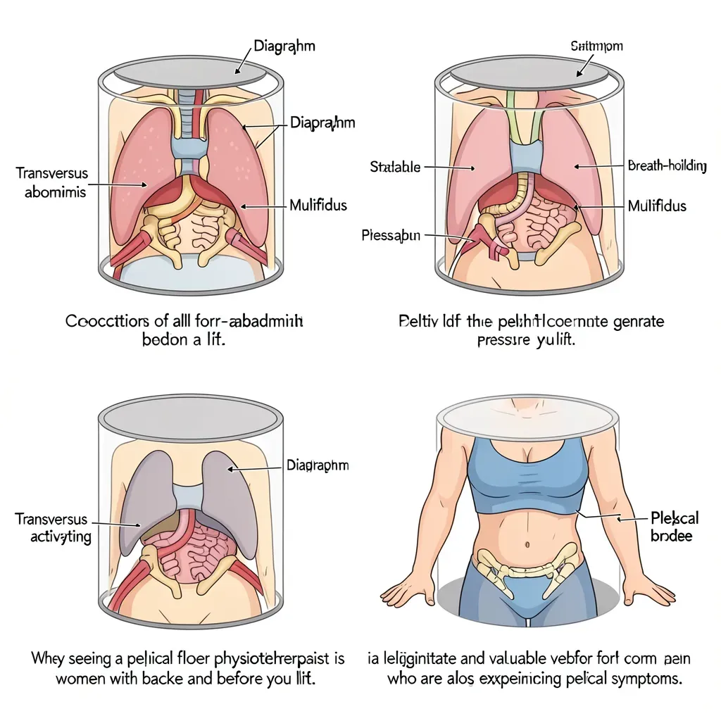
The pelvic floor is the base of the intra-abdominal pressure canister — the system of muscles that, when co-activated, creates the spinal stability required for safe movement. The canister has four walls: the diaphragm above, the transversus abdominis and multifidus around the sides and back, and the pelvic floor below. When any component of this system is dysfunctional, the stability system is compromised.
Pelvic floor dysfunction is far more common in women than men, and far more common than typically acknowledged. Pregnancy and vaginal delivery are the most significant risk factors, but pelvic floor dysfunction also occurs in nulliparous women through high-impact sport, chronic straining, and neurological factors. Dysfunction can mean either weakness (inability to contract adequately) or overactivity (inability to relax fully — a hypertonic pelvic floor).
Both states disrupt the stability canister. A weak pelvic floor cannot generate adequate pressure at the base of the canister during loading. A hypertonic pelvic floor is equally problematic because a muscle that cannot release cannot properly co-activate with the other canister components.
If lumbar stabilisation exercises — particularly the Big 3 — produce worsening of pain or pelvic symptoms, assessment by a pelvic floor physiotherapist is indicated before continuing. The pelvic floor component may need to be addressed before the lumbar stabilisation program will be effective.
Hip Anatomy and Lumbar Loading Patterns
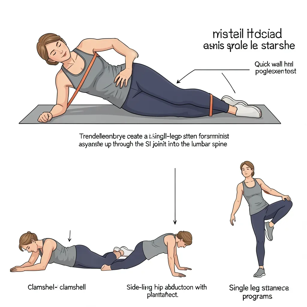
Female pelvic anatomy differs from male anatomy in ways that have direct consequences for lumbar loading. A wider pelvis produces a greater Q angle at the knee, which increases the tendency toward knee valgus (inward collapse of the knee) and femoral internal rotation during weight-bearing activities. This altered lower limb mechanics changes the way forces are transmitted up through the hip into the lumbar spine.
Women also have a higher prevalence of hip pathology — including hip labral tears and femoroacetabular impingement — that can produce referred pain to the groin and anterior hip that mimics lumbar pathology, and vice versa. The hip-spine relationship is clinically important: hip mobility deficits (particularly hip internal and external rotation) are a common but underassessed driver of lumbar overload, as the lumbar spine compensates for restricted hip movement.
Hip mobility assessment and targeted hip strengthening — particularly of the gluteus medius and deep hip rotators — are often underemphasised in female back pain rehabilitation and can produce significant results when included.
If standard lumbar rehabilitation is producing limited improvement, hip mobility and hip abductor/external rotator strength assessment is warranted. Weak gluteus medius and deep hip rotators are disproportionately common in women and can create a compensatory lumbar loading pattern that perpetuates low back symptoms even when the lumbar rehabilitation itself is correct.
The Care Burden and Physical Injury Risk
Women disproportionately perform physical caring roles — primary childcare, aged care for parents, and unpaid household labour. The specific physical demands of caring are among the highest-risk for spinal injury: lifting children from low surfaces (car seats, cots, the floor), carrying children on one hip for extended periods, handling elderly relatives who have limited mobility, and managing domestic tasks in awkward postures.
A mother lifting a toddler from the floor 20-30 times per day is accumulating significant disc loading. Unlike occupational manual handling, where workplace health and safety obligations mandate technique training and equipment provision, domestic care tasks are performed without instruction, without equipment, and without acknowledgement that they carry injury risk.
Practical principles for high-frequency care lifting:
- Hip hinge to the load before lifting — do not round the spine
- Bring the child to you before lifting, reducing the moment arm
- Alternate carry arm when transporting children on the hip
- Use equipment wherever available (change table height, car seat position adjustment)
The Pain Reporting Discrepancy
Research consistently demonstrates that women's pain reports are more likely to be attributed to psychological rather than physical causes by healthcare providers, leading to longer diagnostic delays, less frequent imaging, and less aggressive treatment. This is not a perception — it is a documented phenomenon with real consequences.
Women are more likely to receive antidepressants than analgesia for pain presentations, more likely to be diagnosed with anxiety or depression as the primary problem when pain is the presenting complaint, and less likely to be referred for specialist assessment at equivalent symptom severity compared to men.
Being aware of this disparity is not about cultivating adversarial relationships with healthcare providers. It is about advocating clearly for assessment: documenting symptoms systematically, requesting specific investigations where appropriate, seeking second opinions when the explanation offered does not match the experience, and understanding that the persistence required to obtain an accurate diagnosis is sometimes necessary.
In Review
- Oestrogen receptors in disc and ligament tissue mean hormonal fluctuations across the menstrual cycle directly affect joint laxity and pain threshold — cyclical worsening of back pain before menstruation has a physiological basis.
- Endometriosis causes back pain in a significant proportion of the 10% of reproductive-age women it affects; the diagnostic delay averages 7-10 years, and cyclical pattern, pelvic pain, and dysmenorrhoea are key clinical indicators.
- The pelvic floor is the base of the spinal stability canister; both weakness and hypertonic dysfunction disrupt lumbar stability, and pelvic floor physiotherapy assessment is indicated when standard rehabilitation produces limited results.
- Wider female pelvic anatomy creates a higher Q angle and greater tendency toward femoral internal rotation, altering lumbar loading mechanics; hip abductor and deep rotator strength is frequently underassessed and undertreated.
- Women carry disproportionate physical caring burdens with high-frequency awkward lifting that carries significant disc loading risk; hip hinge technique applied to all care lifting is the primary corrective.
- Women's pain is statistically more likely to be attributed to psychological causes and managed with less diagnostic rigour; systematic symptom documentation and persistent advocacy are sometimes required to obtain accurate assessment.
- Pregnancy-related back pain and pelvic girdle pain are covered in detail in Chapters 28 and 29.