Understanding Disc Herniation
The anatomy of a disc herniation — types, how they happen, natural resorption timeline, and what your MRI actually means.
Disc Anatomy: What You're Working With
The intervertebral disc is not a simple cushion. It's a pressurized hydraulic structure designed to transmit load, allow motion, and protect the spinal cord simultaneously.
Nucleus pulposus: The gel core. Roughly 80% water in youth, gradually losing hydration with age. Under compression it behaves like a pressurized fluid, distributing load outward in all directions.
Annulus fibrosus: The outer wall. Twelve to twenty concentric rings of collagen fibers, each layer oriented at alternating angles (roughly 30 degrees off vertical, alternating direction per layer). This cross-ply arrangement resists both tension and torsion — the same engineering principle used in radial tires.
Cartilaginous end plates: The top and bottom interface between disc and vertebral body. Thin, semi-permeable structures that allow nutrient diffusion into the avascular disc. Their failure is a key initiating event in disc degeneration.
The outer one-third of the annulus is innervated. The inner two-thirds and the nucleus are not. This is why many disc changes produce no pain whatsoever.
Discs have no blood supply. They receive nutrients entirely through diffusion from the end plates — driven by the compression-and-decompression cycle of movement. This is one reason why prolonged immobility accelerates disc degeneration, and why movement is medicine for disc health.
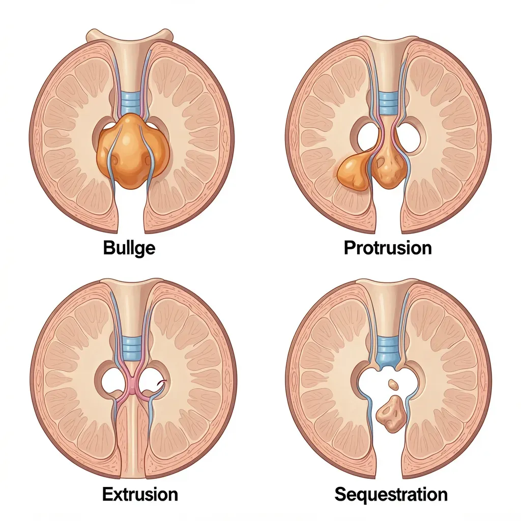
Classification: Bulge, Protrusion, Extrusion, Sequestration
Radiologists use precise terminology that is worth understanding:
Bulge: The disc extends beyond the vertebral margin symmetrically, involving more than 180 degrees of circumference. The annular fibers are intact but bowing outward under pressure. Often normal — found in completely asymptomatic people.
Protrusion: The nucleus has migrated outward but remains contained within the outer annular fibers. The base of the protrusion (measured at the disc margin) is wider than the displaced portion. Still a "contained" herniation.
Extrusion: Nuclear material has pushed through the outer annular fibers and into the spinal canal. The displaced material is now broader than its connection point to the disc — like a mushroom through a hole. Nerve contact is common.
Sequestration (free fragment): The extruded material has completely separated from the disc and floats freely in the spinal canal. No connection remains. This is the most "severe" classification — and often the fastest to resolve, for reasons explained below.
How Herniations Actually Happen
The common narrative — "I lifted something wrong and herniated a disc" — is biomechanically inaccurate. Acute herniations from a single event are rare in healthy discs. What actually happens is a cumulative process.
Research by McGill and colleagues established the sequence:
- Repeated flexion cycles (not single events) cause annular delamination — the concentric layers begin separating from each other in the posterior-lateral region
- The pressurized nucleus begins tracking outward through the delaminated channel with each subsequent loading cycle
- This process may occur over months or years without symptoms
- One day, a seemingly ordinary movement — picking up a pen, getting out of a car — is the final cycle that completes the breach
The person reports: "I herniated my disc reaching for my coffee cup." The coffee cup had nothing to do with it.
Why posterior-lateral? The anterior longitudinal ligament and the posterior placement of the facets constrain anterior and direct posterior herniation. The posterior-lateral corners are the soft spots — the pathway of least resistance.
The activities most responsible for cumulative annular damage: repeated sit-ups and crunches, sustained slouching, repeated forward bending to the floor (especially under load), and any exercise programming that emphasizes spinal flexion under load. Many popular fitness programs are direct contributors to the exact failure mechanism that produces disc herniation.
Natural Resorption: The Counterintuitive Reality
One of the most important and least-communicated facts in spine medicine: disc herniations resorb spontaneously, and larger herniations resorb faster.
This finding has been replicated across multiple studies. A 2017 meta-analysis of 31 studies covering nearly 1,900 patients found that spontaneous resorption occurred in:
- Sequestered discs: 96% showed resorption
- Extruded discs: 70% showed resorption
- Protrusions: 41% showed resorption
- Bulges: 13% showed resorption
The mechanism: extruded nuclear material, once outside the disc, is recognized as foreign by the immune system. Macrophages infiltrate the fragment and phagocytose (consume) it. The larger the fragment and the more contact it has with the epidural space (where immune cells circulate), the faster this process works.
Timeline: significant resorption typically begins within 4-8 weeks, with substantial reduction visible on MRI by 6-12 months.
If your MRI shows a large herniation with nerve contact and your surgeon is recommending urgency — ask them about the resorption timeline. A large extrusion that is not causing progressive neurological deficit (worsening weakness, loss of bowel or bladder control) has an excellent chance of resolving on its own within 12 months. Time, mechanics correction, and rehabilitation are the first-line intervention for the vast majority of herniations.
The 1994 NEJM Study and What Your MRI Means
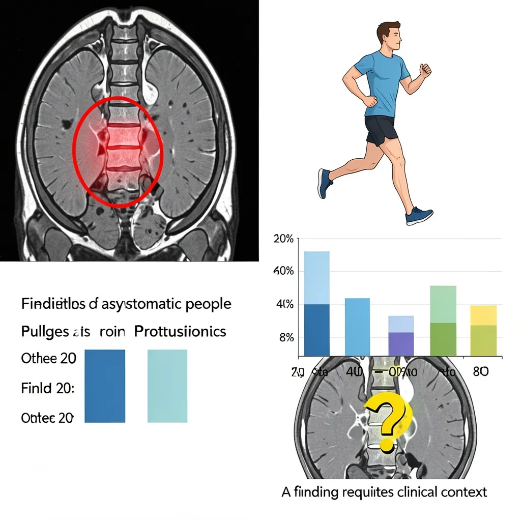
In 1994, the New England Journal of Medicine published a landmark study by Jensen et al. that changed how researchers think about disc findings. They performed MRIs on 98 asymptomatic adults — people with no back pain, no history of back problems, no symptoms whatsoever.
The findings were striking:
- 52% had a bulge at one or more levels
- 27% had a protrusion
- 1% had an extrusion
- Only 36% had a completely "normal" scan
Subsequent studies have confirmed and extended this. Pooled data from thousands of asymptomatic subjects shows age-graded increases in disc findings that are entirely unrelated to pain.
This is not an argument that disc findings are meaningless — they can be clinically significant. It IS an argument against treating a disc finding on an MRI as an explanation for pain without clinical correlation. The question is not "does this person have a disc bulge?" The question is "does this disc finding match the clinical picture — location, symptom behavior, directional testing?"
Level-Specific Patterns
L4-L5: The most common herniation level. The L5 nerve root is typically affected, producing pain radiating from the buttock into the lateral leg and top of the foot. Weakness may occur in great toe extension (extensor hallucis longus).
L5-S1: The second most common level. S1 nerve root involvement produces pain down the back of the leg and into the sole or lateral foot. Calf weakness and reduced Achilles reflex are possible.
Cervical (C5-C6, C6-C7): Produce arm pain (cervical radiculopathy) rather than leg pain. The same mechanics apply — extension typically provides relief, flexion aggravates. Treatment principles parallel the lumbar approach.
Red flags requiring urgent assessment regardless of herniation type: progressive weakness in the legs (not just pain), saddle anesthesia (numbness in the inner thigh and groin), or any change in bowel or bladder function. These suggest cauda equina involvement — a surgical emergency. The vast majority of herniations do not produce these symptoms, but their presence changes the decision tree entirely.
Why Most Herniations Don't Require Surgery
Surgery for disc herniation produces faster short-term relief. At 12 months, outcomes are equivalent between surgery and well-conducted conservative care. At 24 months and beyond, outcomes are indistinguishable.
The only conditions in which surgery provides a durable advantage over conservative care: progressive neurological deficit not responding to treatment, cauda equina syndrome, and intolerable pain unresponsive to all conservative measures after an adequate trial.
The case for attempting conservative rehabilitation first — including the directional preference approach covered in the next article — is strong for the vast majority of patients.
In Review
- The disc is a pressurized hydraulic structure with an innervated outer third — only the outer layers produce pain
- Herniation classification: bulge → protrusion → extrusion → sequestration, in increasing severity of displacement
- Most herniations result from cumulative flexion cycles, not single acute events
- Larger herniations resorb spontaneously at higher rates — extrusions resorb in ~70% of cases
- Over half of asymptomatic adults have disc findings on MRI; imaging findings must be correlated with clinical presentation
- L4-L5 and L5-S1 are the most common herniation levels, each with characteristic nerve root patterns
- Surgery is rarely necessary — conservative care produces equivalent outcomes at 12-24 months for the majority of cases