Spine Anatomy Deep Dive
A complete guide to the structures of the lumbar spine — vertebrae, discs, facets, ligaments, and muscles — and how each contributes to pain.
Why Anatomy Matters for Recovery
Most people with back pain have seen an MRI report or been handed a diagram by a clinician, but they have no framework for interpreting what any of it means. Words like "annular fissure," "ligamentum flavum hypertrophy," or "foraminal narrowing" land without context. Without that context, the natural response is fear — and fear of movement is one of the strongest predictors of prolonged disability.
This chapter builds the anatomical map. It is not a medical school lecture. The goal is functional literacy: enough understanding of what each structure does and how each structure fails that you can interpret your own symptoms, your imaging reports, and the interventions being recommended to you.
The Five Lumbar Vertebrae
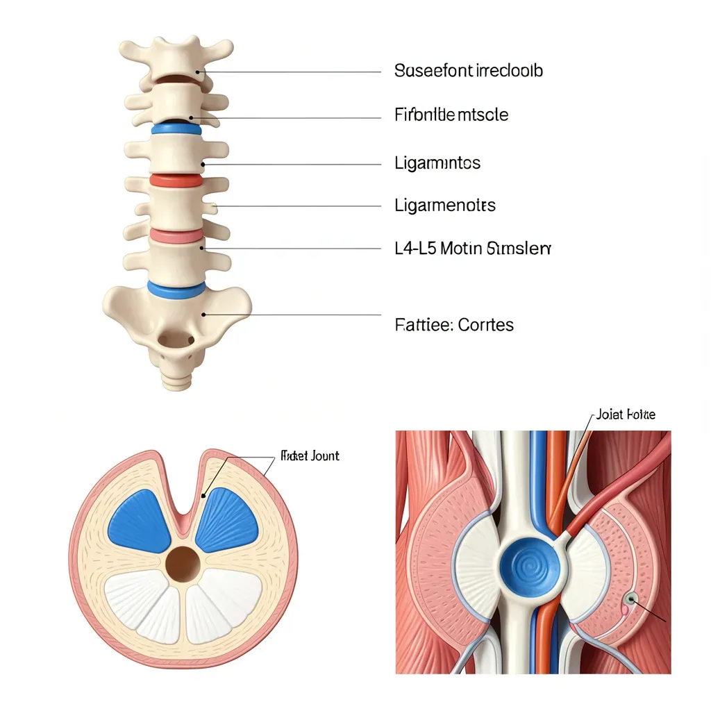
The lumbar spine comprises five vertebrae — L1 through L5 — stacked between the thoracic spine above and the sacrum below. Each vertebra shares the same basic architecture: a large cylindrical body anteriorly, a hollow arch posteriorly (the neural arch), and a series of bony projections — spinous process, transverse processes, and four articular processes — that serve as attachment points for ligaments and muscles and as contact surfaces for the adjacent vertebrae.
The vertebral body is the primary load-bearing structure. It is composed of a hard outer shell (cortical bone) surrounding a porous cancellous interior reinforced by trabecular struts oriented along the primary lines of compressive force. These trabeculae remodel over time in response to loading patterns — a property exploited by rehabilitation when we prescribe specific loading to rebuild bone density and disc health.
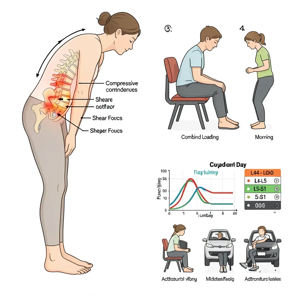
L4-L5 and L5-S1 are the most commonly injured segments for two related reasons. First, they carry the greatest compressive load — the cumulative weight of the upper body concentrates at the bottom of the lumbar column. Second, they exist at a junction between mobile tissue (the lumbar spine) and relatively fixed tissue (the sacrum), creating high shear demands every time the spine flexes or rotates. These junction zones in the body are universally high-risk for injury.
The Intervertebral Disc
The disc sits between each pair of adjacent vertebral bodies. It is not a simple cushion. It is a sophisticated hydraulic and mechanical structure with three distinct layers.
The nucleus pulposus occupies the central core. In a young, healthy disc, it is approximately 80% water — a gel-like material under high internal pressure. This hydrostatic pressure is what gives the disc its load-bearing capacity. Compression from above is distributed radially outward by the pressurized nucleus, rather than transmitted directly through the disc.
The annulus fibrosus surrounds the nucleus as a series of concentric lamellae — roughly 15 to 25 layers of dense collagen fibers. The key architectural feature is that collagen fibers in each lamella run at an angle of approximately 30 degrees to the horizontal, alternating direction between adjacent lamellae. This cross-ply arrangement, similar to steel-belted radial tires, makes the annulus resistant to both compressive and rotational forces. However, it also means that repeated end-range rotation is particularly damaging — it loads the same fiber directions each time, producing cumulative fatigue failure.
The cartilaginous endplates cap the top and bottom of each disc and are the interface between disc tissue and vertebral bone. Their structural integrity is critical for disc health — a fact explored in depth in the next article.
The disc has no direct blood supply after early childhood. All nutrients — glucose, oxygen — must diffuse through the endplates from the vertebral body vasculature. This is why disc health depends so heavily on movement (the pumping mechanism drives diffusion) and why endplate damage is so consequential for long-term disc integrity.
Facet Joints
Each vertebra has two superior and two inferior articular processes that interlock with the adjacent vertebrae, forming the facet joints (also called zygapophyseal joints). There are two facet joints at each spinal level — one on each side.
Facet joints are true synovial joints: they have articular cartilage, a joint capsule, and synovial fluid. Their orientation in the lumbar spine — nearly vertical, facing slightly medially — is designed to resist forward shear and rotation while allowing flexion and extension. This orientation means lumbar facets are stressed primarily in extension and axial rotation.
In the lumbar spine, the facets carry approximately 20% of axial compressive load in normal standing and significantly more in extension. When disc height is lost through degeneration, the facets carry a greater share of load — a progressive transfer that accelerates facet arthritis.
Key Ligaments
The spine's passive stability — the resistance to excessive motion without muscle activation — comes primarily from its ligaments. There are four that matter most for understanding back pain.
The anterior longitudinal ligament (ALL) runs along the front of the vertebral bodies from the skull to the sacrum. It is the strongest spinal ligament and resists hyperextension. It is rarely injured except in severe trauma.
The posterior longitudinal ligament (PLL) runs inside the spinal canal along the posterior aspect of the vertebral bodies. It helps contain disc material that tries to herniate posteriorly. Its relative narrowness in the lumbar region (it fans out in the cervical spine but narrows significantly in the lumbar) explains why posterolateral disc herniations — outside the PLL coverage zone — are far more common than central herniations.
The ligamentum flavum ("yellow ligament") bridges adjacent laminae on the posterior wall of the spinal canal. It is highly elastic and under tension even at rest, which helps maintain the canal's integrity and contributes to spinal stiffness. With age and chronic loading, the ligamentum flavum hypertrophies and may buckle inward, contributing to central stenosis.
The interspinous ligaments connect adjacent spinous processes. They are vulnerable to injury with repeated flexion — particularly the supraspinous ligament (which runs along the tips of the spinous processes). McGill's research on ligament creep showed that sustained flexion postures stretch these ligaments over 20-30 minutes, temporarily reducing their protective stiffness.
Sustained end-range flexion — prolonged sitting slumped forward, sustained forward bending — produces creep deformation in the posterior ligament complex. After 20 minutes in a flexed posture, passive spinal stiffness can decrease by 30-40%, and recovery takes an additional 30-40 minutes of neutral positioning. This is why the first injury of the day often happens not during hard work, but during the routine task performed after a morning of slouched sitting.
The Paraspinal Muscles — Overview
Muscles are the primary stabilizers of the spine during movement. They are covered in depth in Article 41, but the overview here establishes the two-system model important for understanding stability.
The global system — erector spinae, rectus abdominis, external obliques — produces gross trunk movement and transfers load between the thorax and pelvis. These muscles are large, superficial, and generate high force but are not optimized for fine control of intervertebral motion.
The local system — multifidus, transversus abdominis, internal obliques, diaphragm, pelvic floor — controls segmental motion directly. Multifidus, in particular, attaches directly to each lumbar vertebra and monitors and controls the rotational and translational stability of individual segments. After injury, the local system atrophies preferentially and does not spontaneously recover, which is a central reason why McGill's rehabilitation approach targets it specifically.
The Spinal Canal, Foramen, and Neural Structures
The spinal canal runs through the neural arches of stacked vertebrae, housing the spinal cord (which ends at approximately L1-L2 in adults) and the cauda equina below it. The cauda equina is the bundle of nerve roots that continues downward from the cord terminus to exit at each lower lumbar and sacral level.
Nerve roots exit the canal through the intervertebral foramen — openings between adjacent pedicles. Each foramen is bordered superiorly by the pedicle of the vertebra above, inferiorly by the pedicle of the vertebra below, anteriorly by the disc and vertebral body, and posteriorly by the facet joint. This means that disc herniation can compress a nerve root from the front, while facet hypertrophy or ligamentum flavum buckling can compress it from behind.
At L4-L5, the L4 nerve root exits above the disc and the L5 root exits through the foramen bordered by the disc posteriorly. At L5-S1, the S1 root exits through the sacral foramen. This anatomy explains the specific referral patterns for each level of disc herniation — a topic covered in detail in Article 44.
When reading an MRI report, locate the level (L4-L5, L5-S1) and the location within that level (central, paracentral, foraminal, extraforaminal). Central findings are often asymptomatic — the canal is wide enough to tolerate moderate disc material. Foraminal findings are more likely to correlate with symptoms because the nerve root has much less room to maneuver. Location matters more than severity grade for predicting clinical relevance.
In Review
- The lumbar spine has five vertebrae; L4-L5 and L5-S1 carry the greatest load and sit at the mobile-fixed junction with the sacrum, making them the most injury-prone.
- The intervertebral disc is a hydraulic load-distribution system with a pressurized nucleus, a cross-ply annulus resistant to rotation, and cartilaginous endplates that serve as the nutrient gateway.
- Facet joints are synovial joints that primarily resist shear and rotation; they carry 20% of axial load and more in extension, making them vulnerable when disc height is lost.
- The posterior longitudinal ligament narrows in the lumbar region, explaining the predominance of posterolateral herniations.
- Sustained flexion produces ligament creep — a 30-40% reduction in passive stiffness that takes 30-40 minutes to recover — making the hour after prolonged sitting a high-risk window for injury.
- The two-muscle systems model — global movers vs. local segmental stabilizers — explains why the multifidus, not the erector spinae, is the primary rehabilitation target.
- Nerve root compression at the foramen can come from the disc anteriorly or the facet/ligamentum flavum posteriorly; the anatomy of each level determines which roots are affected.